Comparative Study
. 2006 Apr 19;26(16):4155-65.
doi: 10.1523/JNEUROSCI.0175-06.2006.

Synaptic basis for whisker deprivation-induced synaptic depression in rat somatosensory cortex

Affiliations
Comparative Study

Synaptic basis for whisker deprivation-induced synaptic depression in rat somatosensory cortex

Kevin J Bender et al. J Neurosci. .

Abstract

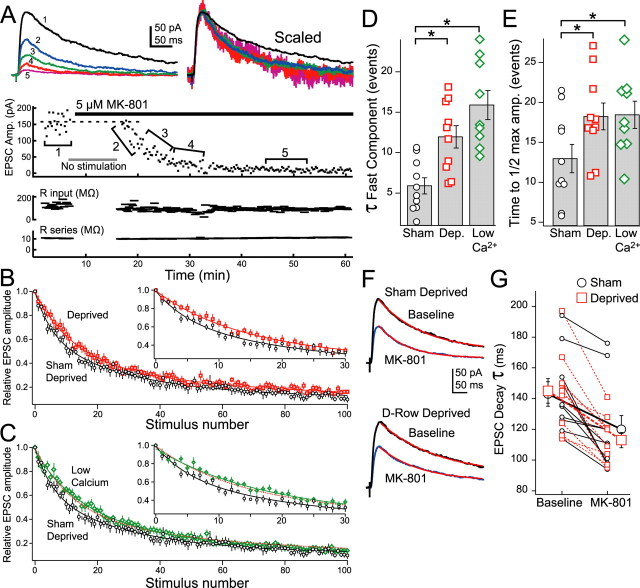
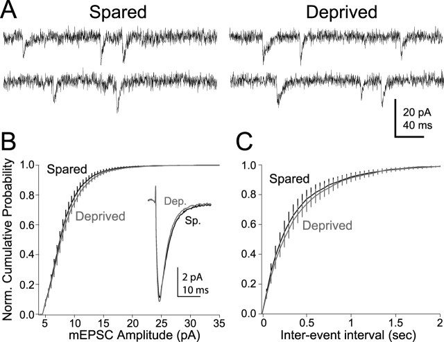
Whisker deprivation weakens excitatory layer 4 (L4) inputs to L2/3 pyramidal cells in rat primary somatosensory (S1) cortex, which is likely to contribute to whisker map plasticity. This weakening has been proposed to represent long-term depression (LTD) induced by sensory deprivation in vivo. Here, we studied the synaptic expression mechanisms for deprivation-induced weakening of L4-L2/3 inputs and assessed its similarity to LTD, which is known to be expressed presynaptically at L4-L2/3 synapses. Whisker deprivation increased the paired pulse ratio at L4-L2/3 synapses and slowed the use-dependent block of NMDA receptor currents by MK-801 [(5S,10R)-(+)-5-methyl-10,11-dihydro-5H-dibenzo[a,d]cyclohepten-5,10-imine maleate], indicating that deprivation reduced transmitter release probability at these synapses. In contrast, deprivation did not alter either miniature EPSC amplitude in L2/3 neurons or the amplitude of quantal L4-L2/3 synaptic responses measured in strontium, indicating that postsynaptic responsiveness was unchanged. In young postnatal day 12 (P12) rats, at least 4 d of deprivation were required to significantly weaken L4-L2/3 synapses. Similar weakening occurred when deprivation began at older ages (P20), when synapses are mostly mature, indicating that weakening is unlikely to represent a failure of synaptic maturation but instead represents a reduction in the strength of existing synapses. Thus, whisker deprivation weakens L4-L2/3 synapses by decreasing presynaptic function, similar to known LTD mechanisms at this synapse.


Figures

Figure 1.
Figure 1.
Time course for deprivation-induced depression of L4-L2/3 synapses. A, Arrangement of large whiskers on rat snout. X, Deprived whiskers. B, Schematic of slice preparation. Barrel columns from whisker rows A and E are labeled. Sites of stimulation, recording, and focal bicuculline (BMI) are shown. C, Representative families of EPSPs at L4-L2/3 synapses recorded in response to increasing L4 stimulation intensity (1–1.5× threshold). Top, Spared column. Bottom, Deprived column of the same slice (5 d of deprivation from P12). D, Input–output curve for EPSP amplitude after 5 d of whisker deprivation. E, Normalized input–output curves after 3, 5, 7, and 9 d of whisker deprivation. Data are normalized to mean EPSP amplitude for cells in spared column at 1.5× threshold. Asterisks indicate deprived columns significantly different from spared columns. F, Identical data from control, nondeprived rats (Allen et al., 2003). G, Time course of deprivation-induced depression of input–output curves, measured by the D:Non-D EPSP ratio (see Results).“Control” and “all 9+” incorporate data from the study by Allen et al. (2003). Numbers in parentheses are cells in the D column. Error bars are SEM.
Figure 2.
Figure 2.
Whisker deprivation increases PPR. A, Top, Representative recordings of pairs of AMPA receptor-mediated EPSCs (recorded at −70 mV) at ISIs of 12, 20, 40, and 80 ms in D and B columns in slices from control and D-row-deprived rats. Bottom, Summary of deprivation effects on PPR. Numbers indicate ISI. B, Lowering external calcium from 2.5 to 1.25 mm increases PPR. C, CTZ does not alter PPR. Veh, DMSO vehicle control. Error bars are SEM.
Figure 3.
Figure 3.
Whisker deprivation slows NMDA-EPSC block by MK-801. A, Example of MK-801-mediated block of transmission. Top left, Averaged NMDAR-mediated EPSC (recorded at +40 mV) in baseline conditions (1) and during MK-801 application (2–5). Top right, EPSCs (2–5) scaled to peak amplitude of 1. Bottom, Individual experiment showing peak EPSC amplitude for each stimulus. B, Average decay in EPSC amplitude for deprived and sham-deprived conditions. Decays are normalized to the amplitude of the first EPSC in MK-801 and fit to double exponentials. C, EPSC decay for sham-deprived and low-calcium conditions. Dashed line, Fit for deprived condition. Error bars are SEM. B, C, Insets, Expansion of stimulus 0–30. D, Time constant for fast component of double exponential decay. E, Time to reach half-maximal EPSC amplitude. Asterisks indicate p < 0.05. F, NMDA-EPSCs before (black) and immediately after (blue) the addition of MK-801, recorded in sham and deprived columns. Red, Single exponential fit of EPSC decay. G, EPSC decay time constant in the absence and presence of MK-801 for sham-deprived (circles) and deprived (squares) conditions. Error bars are SEM.
Figure 4.
Figure 4.
Miniature EPSCs in L2/3 pyramidal cells. A, Representative recordings (at −70 mV) from L2/3 pyramidal neurons in spared (left) and deprived (right) columns. B, Normalized (Norm.) cumulative probability histogram for mEPSC amplitude. Inset, Average mEPSC in deprived and control conditions. C, Normalized cumulative probability histogram for mEPSC interevent interval. Error bars are SEM.
Figure 5.
Figure 5.
Evoked quantal release at L4-L2/3 synapses in the presence of strontium. A, AMPA-mediated EPSC (recorded at −70 mV) at L4-L2/3 synapse in the presence of normal calcium (left). Replacing calcium with strontium produces prolonged release events consisting of individual, quantal events (SrEPSCs). Inset, Average SrEPSC compared with average mEPSC from spared column. B, Relative number (top) and amplitude (amp.; bottom) of SrEPSCs over time window selected for analysis. Time was measured from SrEPSC onset. C, Normalized (Norm.) cumulative probability histogram for SrEPSC amplitude. Inset, Average SrEPSC in deprived and control conditions. D, Average mEPSC and SrEPSC amplitude in deprived and spared conditions. Error bars are SEM.
Figure 6.
Figure 6.
AMPA:NMDA ratio at L4-L2/3 synapses. A, Method for measuring AMPA:NMDA ratio. Traces show mean EPSCs from single representative cells at −80 and +40 mV. The AMPA:NMDA ratio was defined as peak current at −80 mV (AMPA current; black bar) divided by current at +40 mV, 100 ms from EPSC onset (NMDA current; gray bar). B, Summary of AMPA:NMDA ratios in deprived and sham-deprived conditions. Error bars are SEM.
Figure 7.
Figure 7.
Time course for deprivation-induced depression in more mature animals. A, Normalized (Norm.) input–output curves after 2 and 4–6 d of whisker deprivation beginning at P20. Data are normalized to mean EPSP amplitude (Amp.) for cells in spared column at 1.5 times threshold. The asterisk indicates deprived condition significantly different from spared. B, Magnitude of deprivation-induced depression of input–output curves as calculated by D:Non-D ratio. Error bars are SEM.

Similar articles

Cited by

References

    1. Allen CB, Celikel T, Feldman DE (2003). Long-term depression induced by sensory deprivation during cortical map plasticity in vivo. Nat Neurosci 6:291–299. - PubMed
    1. Armstrong-James M, Fox K, Das-Gupta A (1992). Flow of excitation within rat barrel cortex on striking a single vibrissa. J Neurophysiol 68:1345–1358. - PubMed
    1. Bear MF, Cooper LN, Ebner FF (1987). A physiological basis for a theory of synapse modification. Science 237:42–48. - PubMed
    1. Bender KJ, Rangel J, Feldman DE (2003). Development of columnar topography in the excitatory layer 4 to layer 2/3 projection in rat barrel cortex. J Neurosci 23:8759–8770. - PMC - PubMed
    1. Bender VA, Bender KJ, Braiser DJ, Feldman DE (2006). Two coincidence detectors for spike timing-dependent plasticity in somatosensory cortex. J Neurosci 26:4166–4177. - PMC - PubMed

Publication types

MeSH terms

LinkOut - more resources