Comparative Study
. 2006 Apr 19;26(16):4236-46.
doi: 10.1523/JNEUROSCI.4687-05.2006.

Reversal of brain injury-induced prefrontal glutamic acid decarboxylase expression and working memory deficits by D1 receptor antagonism

Affiliations
Comparative Study

Reversal of brain injury-induced prefrontal glutamic acid decarboxylase expression and working memory deficits by D1 receptor antagonism

Nobuhide Kobori et al. J Neurosci. .

Abstract

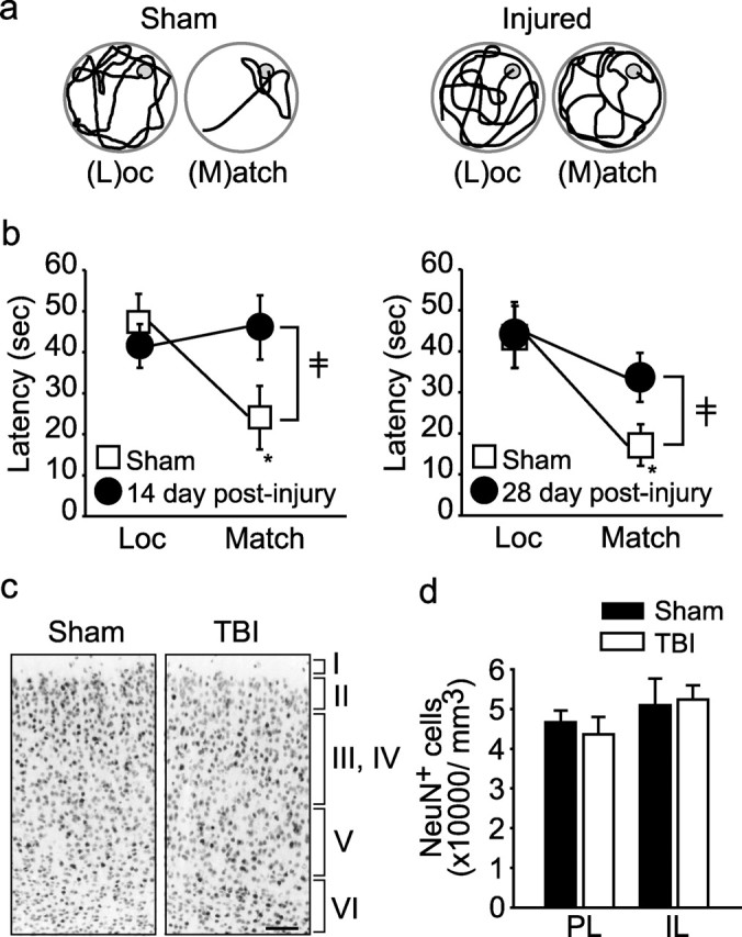
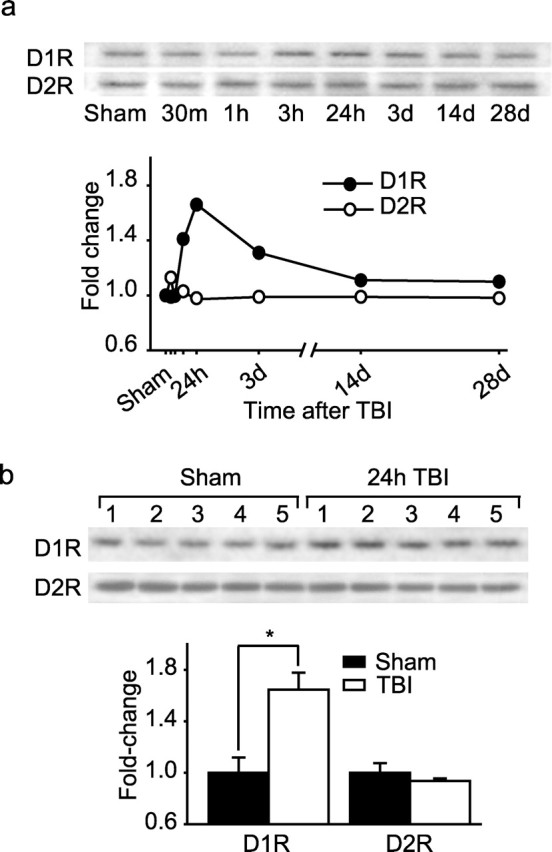
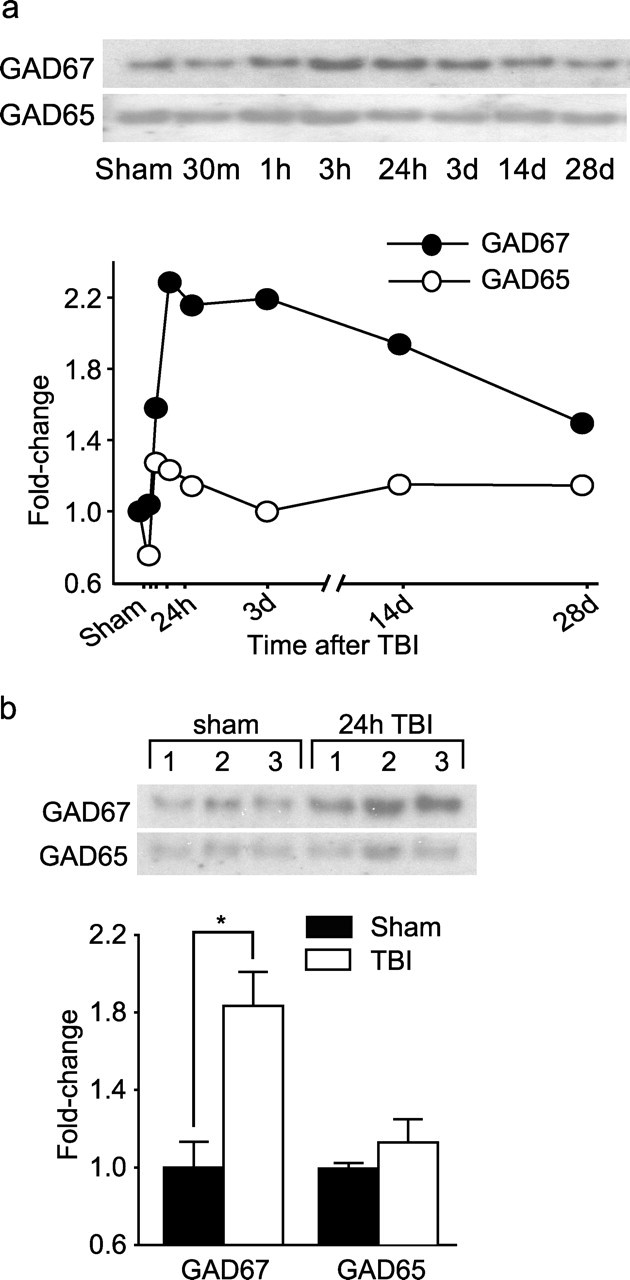
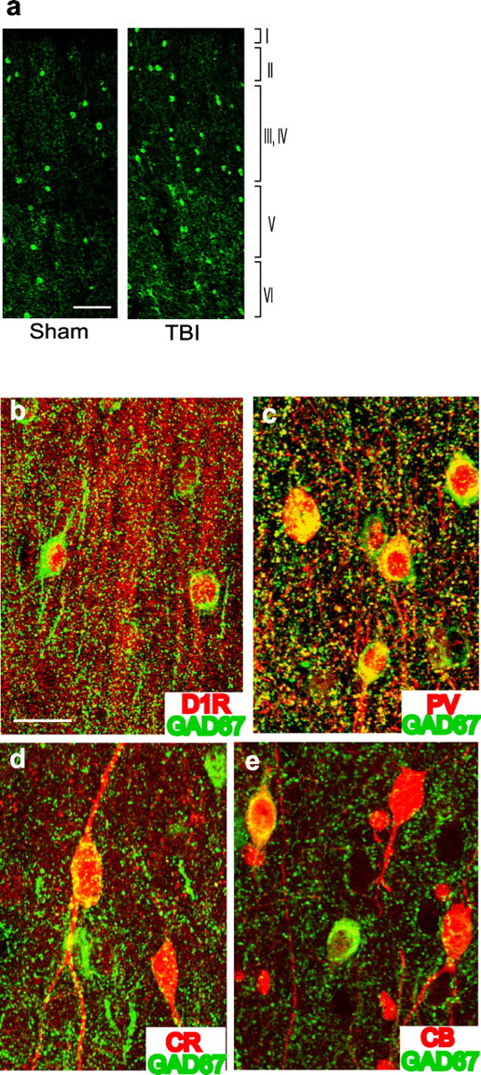
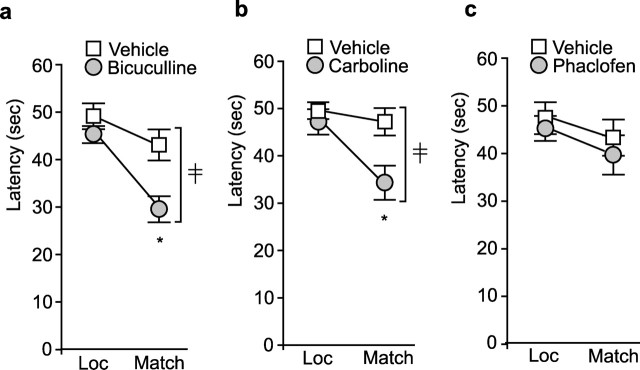
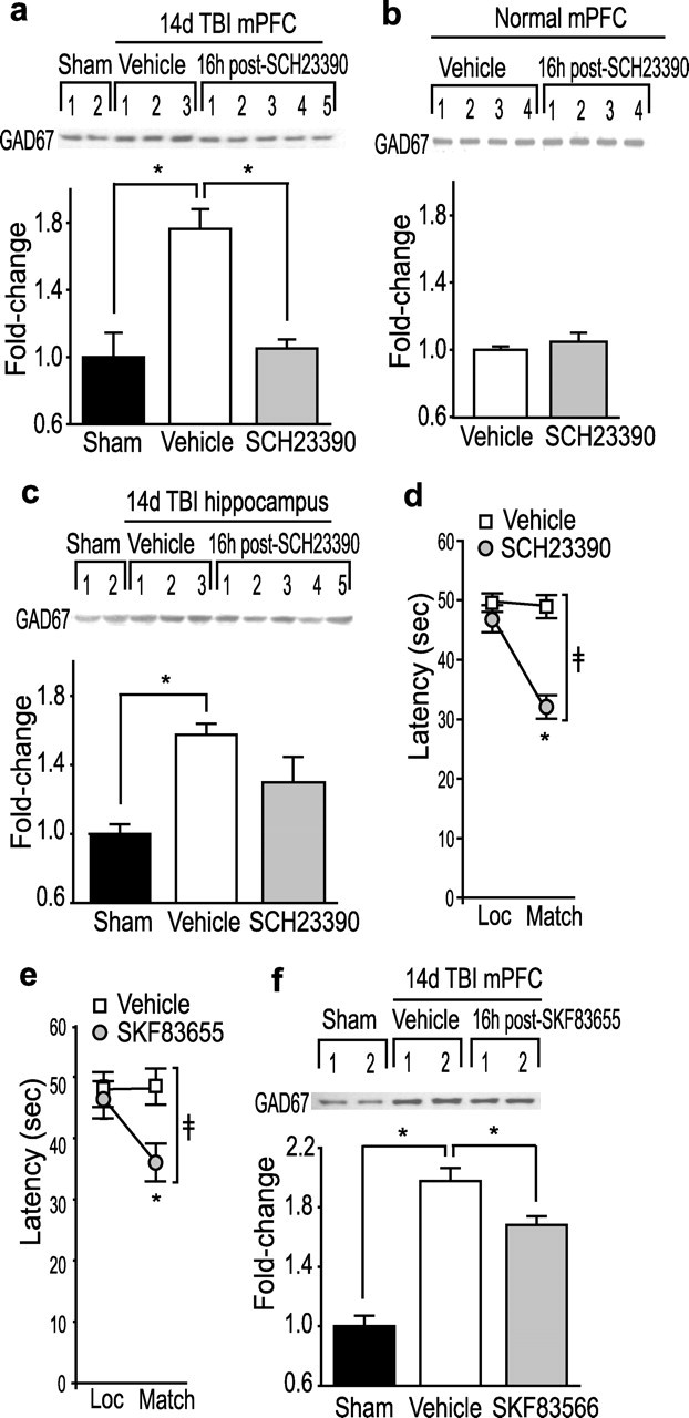
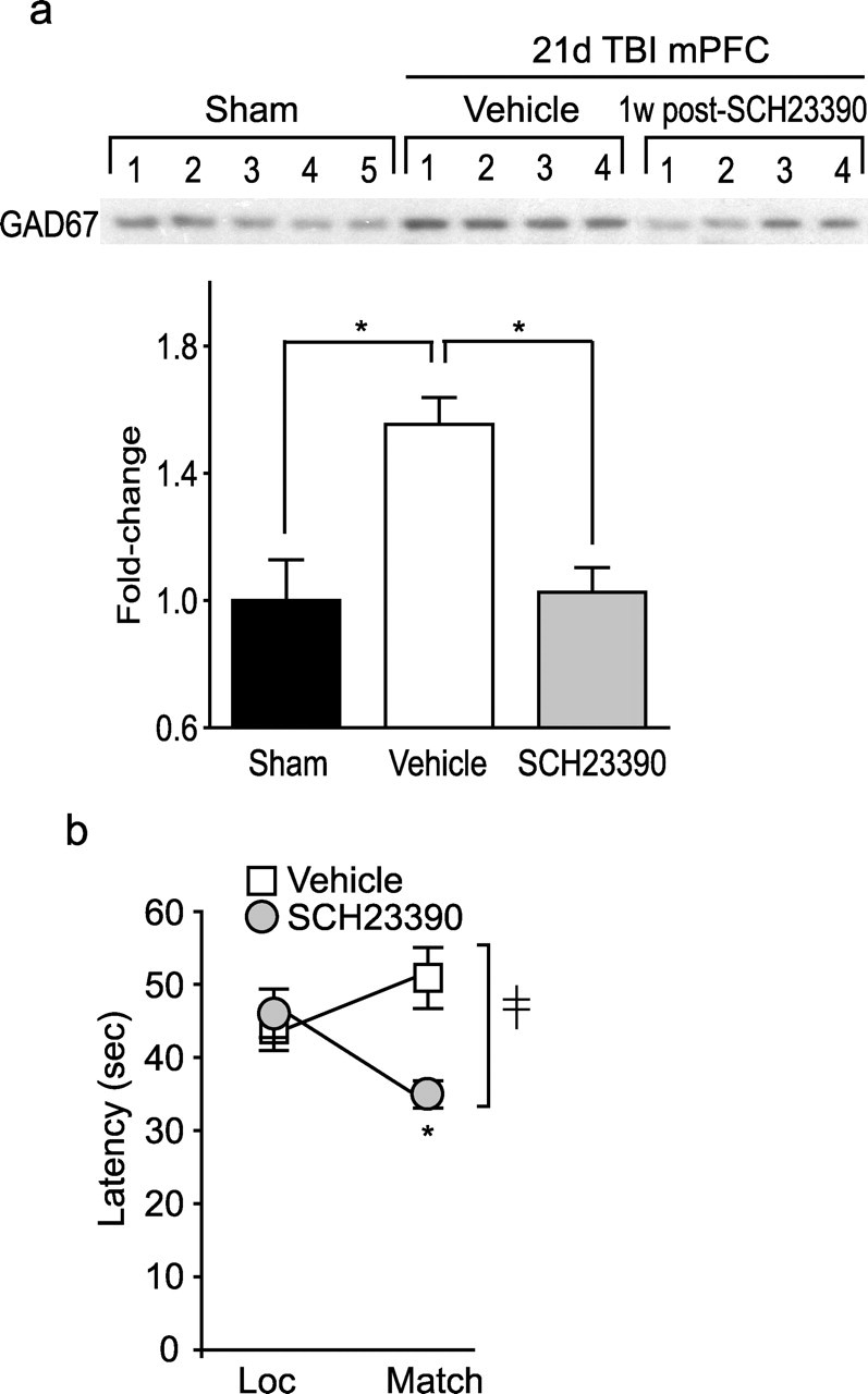
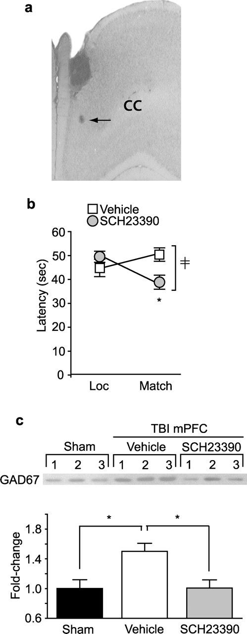
Working memory (WM), the ability to transiently hold information in mind, is essential for high-level cognitive functions that are often impaired in brain-injured patients. The cellular and molecular mechanisms contributing to WM deficits, which can manifest in the absence of overt damage, in these patients are unknown. The function of the dorsolateral prefrontal cortex in humans and monkeys, and the medial prefrontal cortex (mPFC), in rodents is critical for WM. We demonstrate that controlled cortical impact injury of rats causes a long-lasting WM impairment that is associated with increased levels of the GABA-synthesizing enzyme glutamic acid decarboxylase 67 (GAD67) in the mPFC for up to 1 month after injury. A single administration of dopamine D1 antagonists at 14 d after injury is sufficient to decrease GAD67 levels and restore WM for at least 1 week. These findings indicate that inhibition of prefrontal neuronal activity contributes to WM deficits and that strategies to reduce GAD67 expression can offer prolonged WM improvement in brain-injured patients.


Figures

Figure 1.
Figure 1.
Cortical impact injury impairs spatial working memory performance. a, Representative traces of the swimming paths of a sham and an injured rat (14 d after injury) during the location (Loc) and match trials of the delay match-to-place task. b, Summary data displaying average latency to the hidden platform (in seconds) for location trials and match trials during the working memory task for sham and injured rats at 14 d (left) and 28 d (right) after injury (n = 8 per each time point). Significant interaction between trial number and injury status (14 d sham vs injured, F(1,14) = 14.02; 28 d sham vs injured, F(1,14) = 5.299); *significant difference in latency between location and match trial (p < 0.001 in both time points). c, Representative photomicrographs for NeuN-positive neurons in PL/IL region of the mPFC of a sham and a 28 d post-injury animals. Neuronal layers in the mPFC are indicated by Roman numerals. Scale bar, 100 μm. d, Unbiased cell counts from sham (n = 5) and 28 d post-injury (n = 5) animals.
Figure 2.
Figure 2.
Cortical impact injury transiently increases dopamine D1, but not D2, receptor immunoreactivity in the PL/IL cortices. a, Representative Western blots and optical density measurement showing temporal changes in D1 and D2 receptor immunoreactivities in PL/IL protein extracts of sham (24 h) and injured animals at various time points after injury (n = 2 per each time point). b, Representative Western blots and optical density measurements of D1R and D2R immunoreactivities in mPFC of sham (n = 5) and 24 h post-injury (n = 5) animals. ∗p = 0.006.
Figure 3.
Figure 3.
Cortical impact injury increases GAD67, but not GAD65, immunoreactivities in the PL/IL cortices. a, Representative Western blots and optical density measurements of sham (24 h) and injured animals at various time points after injury (n = 2 per each time point). b, Representative Western blots and optical density measurement of GAD67 and GAD65 immunoreactivity in the PL/IL cortices of sham (n = 5) and 24 h post-injury (n = 5) animals. ∗p = 0.019.
Figure 4.
Figure 4.
Increased number of GAD67-positive neurons in the mPFC after brain injury. a, Representative photomicrographs for GAD67-positive cells in mPFC tissue sections of sham and 14 d post-injury animals. Scale bar, 100 μm. Confocal micrographs showing double immunostaining of mPFC tissue section at 14 d after injury with GAD67 and D1R (b), GAD67 and PV (c), GAD67 and CR (d), and GAD67 and CB (e). Scale bar, 20 μm.
Figure 5.
Figure 5.
Systemic administration of GABAA receptor antagonists to injured animals on day 14 improves working memory performance. Summary data showing the latency to locate the hidden platform in the location (Loc) and match trials 30 min after systemic injection of the GABAA antagonist bicuculline (0.5 mg/kg) (a), the benzodiazepine inverse agonist carboline (0.5 mg/kg) (b), and the GABAB antagonist phaclofen (0.5 mg/kg) (c) compared with vehicle-treated injured animals (n = 10 per each drug). Significant interaction between trial number and treatment (F(1,18) = 6.244 for bicuculline; F(1,18) = 8.765 for carboline). ∗Significant difference in latency between location and match trial (p < 0.001 for both bicuculline and carboline).
Figure 6.
Figure 6.
A single administration of D1-selective antagonists decreases mPFC GAD67 immunoreactivity and improves working memory performance in injured animals. a, A representative Western blot and summary results for GAD67 immunoreactivity in mPFC, measured 16 h after a single administration of the D1 antagonist SCH23390 (0.5 mg/kg) or vehicle (n = 10 per group) to injured or sham animals on day 14. ∗p = 0.006 for sham versus TBI vehicle and p < 0.001 for TBI vehicle versus TBI SCH23390. b, A representative Western blot and summary results for GAD67 immunoreactivity in mPFC of the uninjured animals at 16 h after an injection of SCH23390 (0.5 mg/kg) or vehicle (n = 5 per group). c, A representative Western blot and optical density measurement of GAD67 immunoreactivity in the hippocampus, measured 16 h after a single systemic injection of SCH23390 (0.5 mg/kg) or vehicle (n = 5 per group) to injured animals on day 14 after injury. ∗p < 0.001 for sham versus TBI vehicle. d, Summary data show the latency for the injured animals to find the hidden platform in the location (Loc) and match trials at 16 h after systemic injection of SCH23390 (0.5 mg/kg) compared with vehicle treatment (n = 10 per group). Significant interaction between trial number and treatment (F(1,18) = 21.93). ∗Significant difference in latency between location and match trial (p < 0.001). e, Summary data show the latency for the injured animals to find the hidden platform in the location and match trials at 16 h after systemic injection of SKF83655 (50 μg/kg) compared with vehicle treatment (n = 11 per group). Significant interaction between trial number and treatment (F(1,20) = 4.445). ∗Significant difference in latency between location and match trial (p = 0.021). f, A representative Western blot and summary results for GAD67 immunoreactivity in mPFC, measured 18–20 h after a single administration of the D1 antagonist SKF83655 (50 μg/kg) or vehicle (n = 11 per group) to injured or sham animals on day 14. ∗p = 0.011.
Figure 7.
Figure 7.
Lasting effect of D1 receptor antagonist on working memory performance and GAD67 levels. a, Representative Western blot and optical density measurement of GAD67 immunoreactivity in mPFC of sham and injured animals on day 21 after the completion of working memory testing (n = 5 per group). ∗p = 0.004 for sham versus TBI vehicle and p < 0.001 for TBI vehicle versus TBI SCH23390. b, Working memory performance (n = 8 per group) tested 7 d after a single administration of SCH23390 (0.5 mg/kg) on day 14 after injury. Significant interaction between trial number and treatment (F(1,14) = 9.096). ∗Significant difference in latency between location (Loc) and match trial (p = 0.022).
Figure 8.
Figure 8.
Intra-mPFC administration of SCH23390 improves working memory performance and decreases GAD67 level in injured animals. a, Photomicrograph showing the infusion needle track. Arrow, Dye injected through the infusion needle, showing the position of the needle tip. CC, Corpus callosum. b, Summary data of working memory performance 16 h after the targeted infusion of SCH23390 (6 ng/side) or vehicle (n = 9 per group). Significant interaction between trial number and treatment (F(1,16) = 8.325). ∗Significant difference in latency between location (Loc) and match trial (p = 0.014). c, Representative Western blot and optical density measurement of GAD67 immunoreactivity in mPFC of sham and post-injury animals treated with targeted infusion of SCH23390 (6 ng/side) or vehicle (n = 5 per group) after behavioral testing. ∗p = 0.014 for sham versus TBI vehicle and p = 0.006 for TBI vehicle versus TBI SCH23390.

Similar articles

Cited by

References

    1. Akbarian S, Kim JJ, Potkin SG, Hagman JO, Tafazzoli A, Bunney WE Jr, Jones EG (1995). Gene expression for glutamic acid decarboxylase is reduced without loss of neurons in prefrontal cortex of schizophrenics. Arch Gen Psychiatry 52:258–266. - PubMed
    1. Anderson SW, Bechara A, Damasio H, Tranel D, Damasio AR (1999). Impairment of social and moral behavior related to early damage in human prefrontal cortex. Nat Neurosci 2:1032–1037. - PubMed
    1. Arnsten AF (2000). Through the looking glass: differential noradenergic modulation of prefrontal cortical function. Neural Plast 7:133–146. - PMC - PubMed
    1. Asada H, Kawamura Y, Maruyama K, Kume H, Ding R, Ji FY, Kanbara N, Kuzume H, Sanbo M, Yagi T, Obata K (1996). Mice lacking the 65 kDa isoform of glutamic acid decarboxylase (GAD65) maintain normal levels of GAD67 and GABA in their brains but are susceptible to seizures. Biochem Biophys Res Commun 229:891–895. - PubMed
    1. Asada H, Kawamura Y, Maruyama K, Kume H, Ding RG, Kanbara N, Kuzume H, Sanbo M, Yagi T, Obata K (1997). Cleft palate and decreased brain gamma-aminobutyric acid in mice lacking the 67-kDa isoform of glutamic acid decarboxylase. Proc Natl Acad Sci USA 94:6496–6499. - PMC - PubMed

Publication types

MeSH terms