Comparative Study
. 2006 Apr 19;26(16):4277-88.
doi: 10.1523/JNEUROSCI.5078-05.2006.

Beta-amyloid accumulation impairs multivesicular body sorting by inhibiting the ubiquitin-proteasome system

Affiliations
Comparative Study

Beta-amyloid accumulation impairs multivesicular body sorting by inhibiting the ubiquitin-proteasome system

Claudia G Almeida et al. J Neurosci. .

Abstract

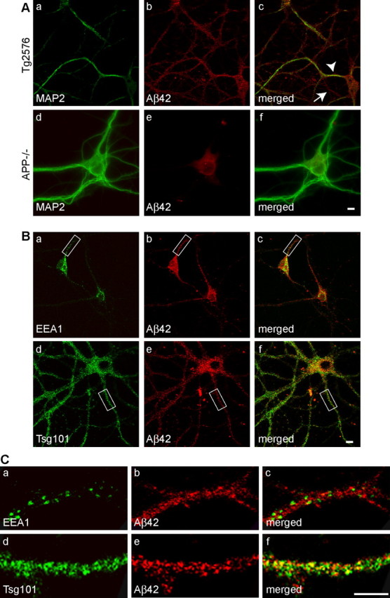
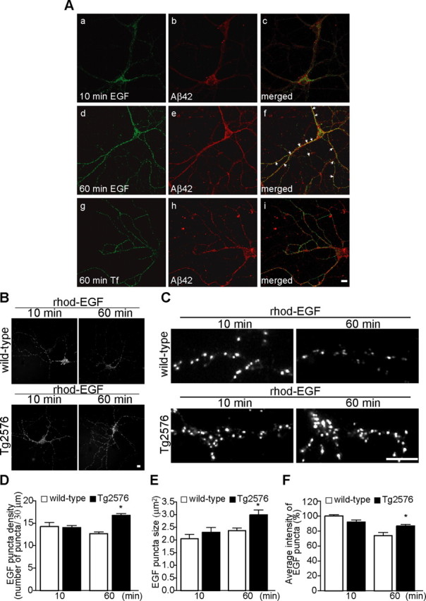
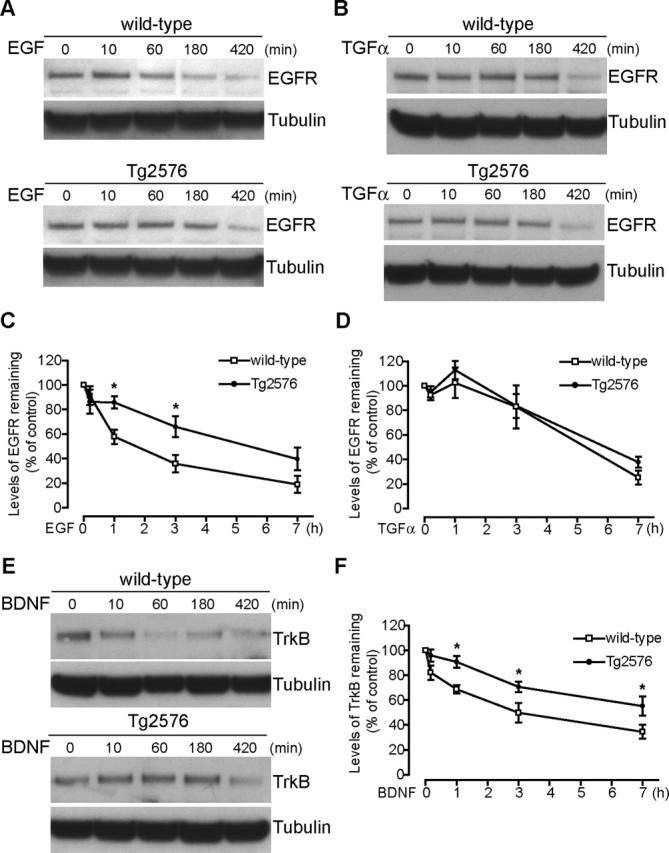
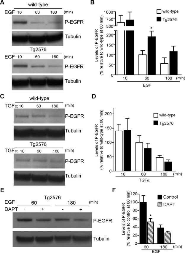
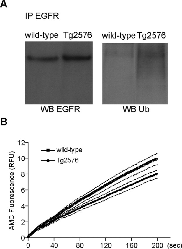
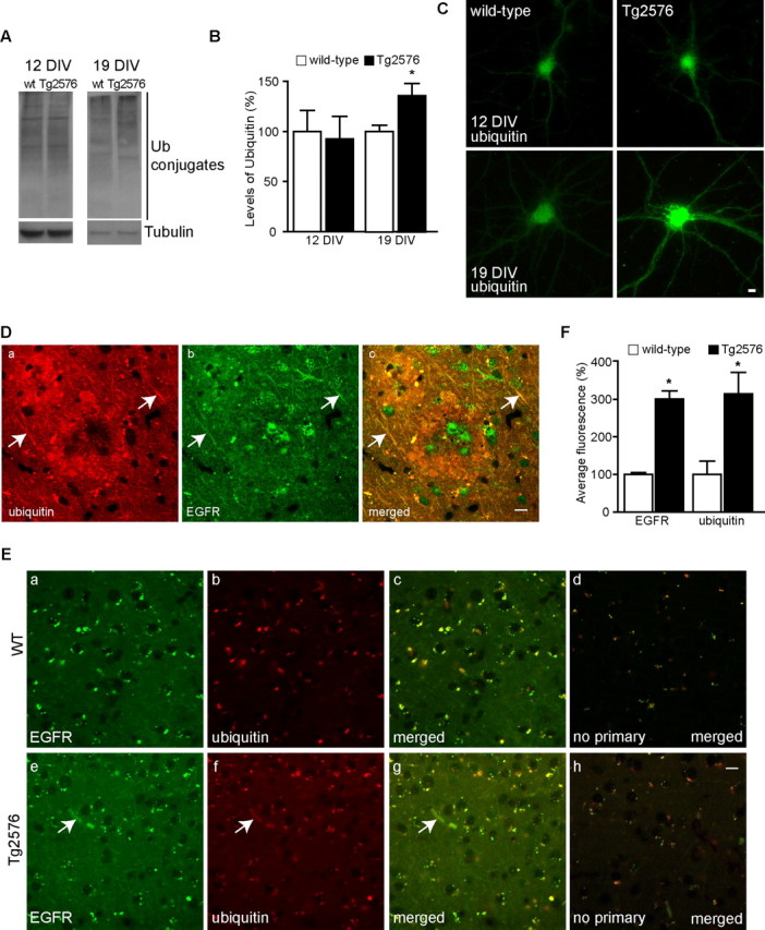
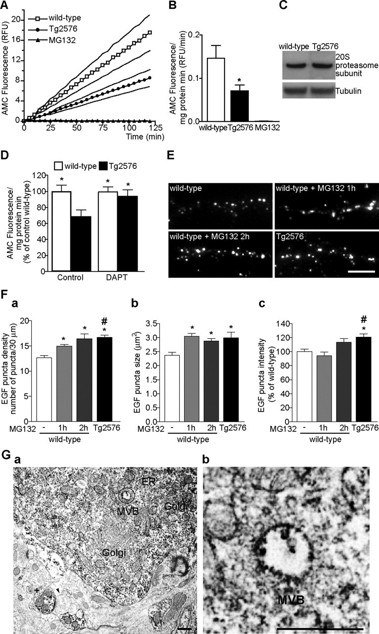
Increasing evidence links intraneuronal beta-amyloid (Abeta42) accumulation with the pathogenesis of Alzheimer's disease (AD). In Abeta precursor protein (APP) mutant transgenic mice and in human AD brain, progressive intraneuronal accumulation of Abeta42 occurs especially in multivesicular bodies (MVBs). We hypothesized that this impairs the MVB sorting pathway. We used the trafficking of the epidermal growth factor receptor (EGFR) and TrkB receptor to investigate the MVB sorting pathway in cultured neurons. We report that, during EGF stimulation, APP mutant neurons demonstrated impaired inactivation, degradation, and ubiquitination of EGFR. EGFR degradation is dependent on translocation from MVB outer to inner membranes, which is regulated by the ubiquitin-proteasome system (UPS). We provide evidence that Abeta accumulation in APP mutant neurons inhibits the activities of the proteasome and deubiquitinating enzymes. These data suggest a mechanism whereby Abeta accumulation in neurons impairs the MVB sorting pathway via the UPS in AD.


Figures

Figure 1.
Figure 1.
Late endocytic localization of Aβ42 in neurites of cultured APP mutant neurons. Neurons from APP mutant transgenic mice harboring the human AD Swedish mutation (Tg2576) were immunostained with an antibody specific for the C terminus of Aβ42 (Chemicon) and other subcellular markers at 19 DIV. Aa–Ac, Aβ42 localized to both dendrites, identified by MAP2-positive staining (arrowhead), and axons (arrow), identified by the absence of MAP2 staining. Ad–Af, Absence of the punctate Aβ42 immunostaining in neurites of APP−/− neurons; there was low level of background fluorescence in the cell body. Ba–Bc, Aβ42 did not colocalize with EEA1, a marker of early endosomes. Bd–Bf, Aβ42 overlap was evident with Tsg101, a marker of MVBs. C, Detail of neurites indicated in B, demonstrating Aβ42 colocalization with Tsg101 (d–f) but not with EEA1 (a–c). Scale bars, 10 μm.
Figure 2.
Figure 2.
EGF endocytosis to the MVB sorting pathway is altered in cultured APP mutant neurons. Wild-type and APP mutant neurons (12 DIV) were incubated with rhod-EGF (500 ng/ml) or Alexa 488–Tf (20 μg/ml) for the indicated times at 37°C. Aa–Ac, rhod-EGF uptake after 10 min incubation followed by Aβ42 staining. Aβ42 did not share compartments with EGF after 10 min internalization, at which time EGF is in the early endocytic pathway. Ad–Af, rhod-EGF uptake after 60 min incubation followed by Aβ42 staining. Aβ42 shared more labeled compartments with EGF after 60 min internalization, at which time EGF traffics to the MVB sorting pathway. Ag–Ai, Alexa 488–Tf uptake after 60 min incubation followed by Aβ42 staining. Aβ42 did not share compartments with Tf after 60 min of uptake, at which time Tf localizes to the recycling pathway. B, Comparison of rhod-EGF endocytosis at 10 and 60 min in neurites of wild-type and APP mutant neurons (representative images). C, Representative higher-magnification images of neurites from wild-type and APP mutant neurons after 10 and 60 min internalization of rhod-EGF (segments of 30 μm). D, Quantification of the number of EGF puncta per 30 μm segment of neurite at 10 and 60 min after rhod-EGF internalization in wild-type (white bar) and APP mutant (red bar) neurons. At 10 min, there was no difference in the number of EGF puncta between wild-type (56 neurites; n = 3) and APP mutant (56 neurites; n = 3) neurons. At 60 min, there was a 33.4 ± 4.4% increase in rhod-EGF puncta density in APP mutant (290 neurites; n = 11) compared with wild-type (408 neurites; n = 15) neurons (∗p < 0.05 relative to wild-type neurons at 60 min). E, Quantification of the size of EGF puncta at 10 and 60 min after rhod-EGF internalization. At 10 min, there was no difference between wild-type (56 neurites; n = 3) and APP mutant (56 neurites; n = 3) neurons. At 60 min, there was a 26.2 ± 8.9% increase in rhod-EGF puncta size in APP mutant (290 neurites; n = 11) compared with wild-type (408 neurites; n = 15) neurons (∗p < 0.05 relative to wild-type neurons at 60 min). F, Quantification of the intensity of EGF puncta at 10 and 60 min after rhod-EGF internalization. In wild-type neurons (138 neurites; n = 3), there was a 25.9 ± 4.4% reduction in intensity of rhod-EGF puncta from 10 to 60 min (∗p < 0.05 relative to wild-type neurons at 10 min), whereas in APP mutant neurons (141 neurites; n = 3), there was no statistically significant reduction in EGF signal between the two time points. Error bars indicate SEM. Scale bars, 10 μm.
Figure 3.
Figure 3.
Delayed degradation of EGFR after EGF, but not TGFα, and of TrkB after BDNF activation, in APP mutant neurons. Wild-type and APP mutant neurons (12 DIV) were incubated with EGF (500 ng/ml), TGFα (500 ng/ml), or BDNF (50 ng/ml) for the indicated times at 37°C, and then cell lysates were subjected to SDS-PAGE and immunoblotted for EGFR or TrkB and for α-tubulin, as a loading control. A, During treatment with EGF, preserved EGFR signal, consistent with reduced degradation, was evident in APP mutant compared with wild-type cell lysates at and after 60 min. B, During treatment with TGFα, no difference in EGFR signal was evident in APP mutant compared with wild-type cell lysates at and after 60 min, consistent with an unaltered recycling pathway. C, Quantification of remaining levels of EGFR relative to unstimulated conditions revealed that EGF activation induced greater downregulation of EGFR in wild-type (open squares; n = 6–16 for each time point) compared with APP mutant (filled circles; n = 6–16 for each time point) neurons, which was statistically significant at 1 and 3 h (∗p < 0.05). D, In contrast, TGFα activation induced a similar downregulation of EGFR in wild-type (open squares; n = 7) and APP mutant (filled circles; n = 7) neurons, consistent with an unaltered TGFα–EGFR recycling pathway. E, Similar to EGFR after EGF treatment, the decrease in levels of TrkB was delayed in APP mutant neurons, consistent with reduced degradation in APP mutant compared with wild-type neurons at and after 60 min of BDNF treatment. F, Quantification of TrkB levels revealed that BDNF activation induced greater downregulation of TrkB in wild-type (open squares; n = 6–10 for each time point) compared with APP mutant (filled circles; n = 6–12 for each time point) neurons, which was statistically significant at 1, 3, and 7 h (∗p < 0.05). Error bars indicate SEM.
Figure 4.
Figure 4.
Delayed dephosphorylation of EGFR after EGF, but not TGFα, treatment in APP mutant neurons. A, After EGF treatment, cell lysates (12 DIV) were subjected to SDS-PAGE and immunoblotted for phospho-EGFR (pY1173-EGFR) and for α-tubulin, as a loading control. The preserved P-EGFR signal in APP mutant compared with wild-type neurons at 60 min is consistent with reduced deactivation of the receptor. B, EGFR deactivation, quantified by reduction in levels of P-EGFR after EGF treatment for 60 and 180 min, was greater in wild-type (white bars; n = 5–9 for each time point) compared with APP mutant (black bars; n = 6–9 for each time point) neurons. Deactivation of EGFR was significantly reduced by 89.6 ± 35.8% in APP mutant neurons after 60 min treatment with EGF (∗p < 0.05). C, In contrast, after TGFα treatment, no difference was detected in P-EGFR signal in APP mutant compared with wild-type neurons. D, Quantification of EGFR deactivation revealed no difference in APP mutant compared with wild-type neurons after TGFα treatment. E, Treatment of APP mutant neurons with the γ-secretase inhibitor DAPT (250 nm, 17 h), before the indicated time points of EGF treatment, reduced the levels of P-EGFR. F, Quantification of EGFR deactivation in APP mutant neurons with or without DAPT treatment. There was a 47.2 ± 17.9% increase in EGFR deactivation (hatched bars; n = 9) in DAPT-treated compared with untreated APP mutant (black bars; n = 8) neurons at 60 min (∗p < 0.05). Error bars indicate SEM.
Figure 5.
Figure 5.
Decreased deubiquitination of EGFR after EGF stimulation in APP mutant neurons. A, Wild-type and APP mutant transgenic neurons (12 DIV) were incubated with EGF (500 ng/ml) for 1 h at 37°C. Cell lysates were subjected to immunoprecipitation (IP) with anti-EGFR, followed by SDS-PAGE and immunoblotting for EGFR or ubiquitin. Representative blots revealed elevated levels of ubiquitinated-EGFR in APP mutant compared with wild-type neurons. B, Kinetics of Ub-AMC hydrolysis, a measure of deubiquitinating enzyme activity, revealed decreased deubiquitination activity in cell lysates from APP mutant (filled circles; n = 9) compared with wild-type (open squares; n = 7) neurons. Data are expressed as mean (solid line) ± SEM (dotted lines). RFU, Relative fluorescence units.
Figure 6.
Figure 6.
Increased ubiquitin and EGFR in APP mutant neurons. A, Cell lysates from 12 and 19 DIV wild-type (wt) and APP mutant neurons (both without EGF treatment) were subjected to SDS-PAGE and immunoblotted for ubiquitin and for α-tubulin, as a loading control. APP mutant neurons revealed a similar pattern of ubiquitin conjugates to wild-type neurons at 12 DIV and an increase in total ubiquitin conjugates at 19 DIV. B, Quantification of levels of ubiquitin conjugates indicated that there was no significant difference at 12 DIV, whereas at 19 DIV, there was a 35.9 ± 12.9% increase in ubiquitin conjugates in APP mutant (black bars; n = 9) compared with wild-type (white bars; n = 12) neurons (∗p < 0.05). C, By immunofluorescence, ubiquitin immunoreactivity was increased especially in processes of APP mutant compared with wild-type neurons at 19 DIV but not at 12 DIV. Da–Dc, Representative tissue section from a late stage (18 months) Tg2576 mouse brain double labeled with anti-EGFR (green) and anti-ubiquitin (red) revealed that increased EGFR and ubiquitin immunoreactivity colocalized in dystrophic neurites (arrows) around a plaque. E, Representative images from cortical areas without plaques from late stage (18 months) Tg2576 (e–h) and wild-type (a–d) mice. Anti-EGFR (a, e, green), anti-ubiquitin (b, f, red), merged image (c, g), and secondary antibodies (d, h) in the absence of primary antibodies (merged image). Tg2576 brain sections showed an increase in immunoreactivity for EGFR and ubiquitin when compared with wild-type. F, Quantification of total fluorescence after subtraction of background staining showed an almost threefold increase in EGFR and ubiquitin immunoreactivity in Tg2576 as compared with wild-type brain (∗p < 0.05; n = 3). Scale bars, 10 μm. Error bars indicate SEM.
Figure 7.
Figure 7.
Aβ-dependent inhibition of the proteasome. A, Proteasome activity was assayed in lysates prepared from wild-type (open squares) and APP mutant (filled circles) neurons (12 DIV). The kinetics of fluorescence accumulation from the degradation of the proteasome substrate Suc-LLVY-AMC monitored for 120 min revealed markedly increased AMC accumulation in wild-type compared with APP mutant neurons. Addition of MG132 completely prevented AMC accumulation (filled triangles). Data are expressed as mean (solid line) ± SEM (dotted lines). B, Bar graph of the average slope obtained by linear regression analysis during the 120 min of Suc-LLVY-AMC degradation in wild-type (white bar; n = 14), APP mutant (black bar; n = 16), and MG132 (black bar; n = 2); there was a 51.4 ± 21.4% decrease in proteasome activity in APP mutant compared with wild-type neurons (∗p < 0.05). C, There was no appreciable difference in the levels of the 20S proteasome α-subunit in wild-type compared with APP mutant neurons (representative blots). D, Treatment of APP mutant neurons with DAPT (250 nm, 17 h; n = 11) normalized the levels of proteasome activity to wild-type (n = 11) levels (∗p < 0.05 relative to untreated APP mutant neurons; n = 9). Treatment with DAPT had no statistically significant effect on proteasome activity in wild-type neurons (n = 11). E, Representative images of neurites from wild-type and APP mutant neurons after 1 h internalization of rhod-EGF (segments of 30 μm). Wild-type neurons pretreated with MG132 for 1 or 2 h revealed similar EGF uptake to that in APP mutant neurons. Scale bar, 10 μm. Fa, The bar graph depicts increased number of rhod-EGF puncta in wild-type neurons treated with MG132 (112 neurites, n1 h = 7; 119 neurites, n2 h = 5), especially after 2 h (n = 5) to levels similar to that in APP mutant neurons (290 neurites, n = 11) [∗p < 0.05 relative to untreated wild-type neurons (408 neurites, n = 15); #p < 0.05 relative to wild-type neurons pretreated for 1 h with MG132 (112 neurites, n = 7)]. Fb, The size of EGF puncta in wild-type neurons treated with MG132 was significantly increased only after 1 h, reaching APP mutant levels (∗p < 0.05). Fc, Quantification of the intensity of rhod-EGF puncta revealed that 2 h of treatment with MG132 was necessary to increase rhod-EGF puncta intensity. Error bars indicate SEM. Ga, Immunoreactivity of the β-subunit of the 20S proteasome in normal rat brain was evident in the cytoplasm of pyramidal neurons and in dendrites (+). Immunoperoxidase labeling was distributed diffusely in neuronal perikaryon, with pronounced labeling especially associated with MVBs, endoplasmic reticulum, and small vesicles. In contrast, Golgi revealed little labeling. A presynaptic compartment (arrowhead) had no appreciable labeling, whereas the postsynaptic compartment to which it is connected by a synaptic density demonstrates marked staining. Gb, Higher-magnification image of circled area in a revealed labeling in the outer membrane and inner vesicle of a MVB. ER, Endoplasmic reticulum; Golgi, Golgi apparatus; RFU, relative fluorescence units. Scale bars, 500 nm.

References

    1. Almeida CG, Tampellini D, Takahashi RH, Greengard P, Lin MT, Snyder EM, Gouras GK (2005). Beta-amyloid accumulation in APP mutant neurons reduces PSD-95 and GluR1 in synapses. Neurobiol Dis 20:187–198. - PubMed
    1. Alwan HA, van Zoelen EJ, van Leeuwen JE (2003). Ligand-induced lysosomal epidermal growth factor receptor (EGFR) degradation is preceded by proteasome-dependent EGFR de-ubiquitination. J Biol Chem 278:35781–35790. - PubMed
    1. Babst M (2005). A protein’s final ESCRT. Traffic 6:2–9. - PubMed
    1. Bertram L, Tanzi RE (2005). The genetic epidemiology of neurodegenerative disease. J Clin Invest 115:1449–1457. - PMC - PubMed
    1. Bertram L, Hiltunen M, Parkinson M, Ingelsson M, Lange C, Ramasamy K, Mullin K, Menon R, Sampson AJ, Hsiao MY, Elliott KJ, Velicelebi G, Moscarillo T, Hyman BT, Wagner SL, Becker KD, Blacker D, Tanzi RE (2005). Family-based association between Alzheimer’s disease and variants in UBQLN1. N Engl J Med 352:884–894. - PubMed

Publication types

MeSH terms