Comparative Study
. 2006 Apr 19;26(16):4289-97.
doi: 10.1523/JNEUROSCI.4178-05.2006.

Compartment-dependent colocalization of Kir3.2-containing K+ channels and GABAB receptors in hippocampal pyramidal cells

Affiliations
Comparative Study

Compartment-dependent colocalization of Kir3.2-containing K+ channels and GABAB receptors in hippocampal pyramidal cells

Akos Kulik et al. J Neurosci. .

Abstract

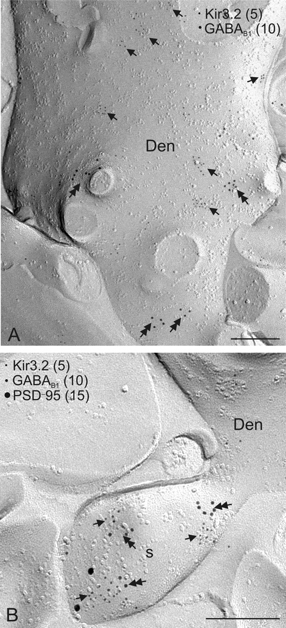
G-protein-coupled inwardly rectifying K+ channels (Kir3 channels) coupled to metabotropic GABAB receptors are essential for the control of neuronal excitation. To determine the distribution of Kir3 channels and their spatial relationship to GABAB receptors on hippocampal pyramidal cells, we used a high-resolution immunocytochemical approach. Immunoreactivity for the Kir3.2 subunit was most abundant postsynaptically and localized to the extrasynaptic plasma membrane of dendritic shafts and spines of principal cells. Quantitative analysis of immunogold particles for Kir3.2 revealed an enrichment of the protein around putative glutamatergic synapses on dendritic spines, similar to that of GABA(B1). Consistent with this observation, a high degree of coclustering of Kir3.2 and GABA(B1) was revealed around excitatory synapses by the highly sensitive SDS-digested freeze-fracture replica immunolabeling. In contrast, in dendritic shafts receptors and channels were found to be mainly segregated. These results suggest that Kir3.2-containing K+ channels on dendritic spines preferentially mediate the effect of GABA, whereas channels on dendritic shafts are likely to be activated by other neurotransmitters as well. Thus, Kir3 channels, localized to different subcellular compartments of hippocampal principal cells, appear to be differentially involved in synaptic integration in pyramidal cell dendrites.


Figures

Figure 1.
Figure 1.
Distribution of immunoreactivity for the Kir3.2 subunit in the hippocampus. A, B, The immunostaining was moderate to strong in dendritic layers of the CA area and dentate gyrus in the rat (A) and wild-type (WT) mouse (B). In the CA1, the immunolabeling for the protein was strong and homogeneous in the stratum lacunosum-moleculare, whereas the strata oriens and radiatum showed uneven immunostaining with moderate intensity of immunoreactivity in the proximal half and high intensity in the distal half of these layers. In CA3, the immunoreactivity for Kir3.2 was strong in the strata oriens, radiatum, and lacunosum-moleculare. In the dentate gyrus, strong immunostaining was detected in the hilus and moderate in the molecular layer. C, No immunoreactivity for Kir3.2 was found in the hippocampus of the Kir3.2-deficient (KO) mice. Scale bars, 200 μm. o, Stratum oriens; p, stratum pyramidale; r, stratum radiatum; l-m, stratum lacunosum-moleculare; m, stratum moleculare; g, stratum granulosum; h, hilus.
Figure 2.
Figure 2.
Preferential postsynaptic localization of Kir3.2 in dendritic layers of the CA1 area. Electron micrographs show immunoreactivity for the Kir3.2 subunit in the strata oriens (A, C), radiatum (B, D, E), and lacunosum-moleculare (>F–I) as detected by the preembedding immunogold method. A, B, F, G, I, Clusters of immunogold particles were seen along the extrasynaptic plasma membrane (arrows) of dendritic shafts (Den) of pyramidal cells contacted by terminals (T) of presumed GABAergic cells. Labeling was occasionally found at the edge of symmetrical synaptic specializations (arrowheads in B, G). A, CF, H, I, Immunoparticles were abundant on the extrasynaptic plasma membrane (arrows) of dendritic spines of pyramidal cells (s). They also appeared occasionally over the postsynaptic specialization (double arrowhead in C) at synapses between axon terminals (T) of putative pyramidal cells and dendritic spines and at the edge of asymmetrical synapses (arrowheads in D, F). C, E, Presynaptically, immunogold particles (double arrows) were localized to the extrasynaptic plasma membrane and to the presynaptic membrane specialization of axon terminals (T) establishing asymmetrical synapses. I, Immunolabeling was also visible in dendritic shafts of presumed interneurons (Den1) establishing asymmetrical synapses with presynaptic boutons (T1, T2). Note that the dendritic spine (s), contacted by an axon terminal (T3), and the dendritic shaft (Den2) of a pyramidal cell are also labeled. Scale bars, 0.2 μm.
Figure 3.
Figure 3.
Distribution of immunoparticles for the Kir3.2 and GABAB1 subunits relative to symmetrical and asymmetrical synapses on dendrites of CA1 pyramidal cells as assessed by preembedding immunogold labeling. A, Histogram showing the spatial distribution of immunoparticles for Kir3.2 (open bars; n = 209) and GABAB1 (filled bars; n = 241) around symmetrical synapses on dendritic shafts. Distances of immunogold particles were measured from the closest edge of the synapses along the surface of dendritic shafts reconstructed from serial ultrathin sections. Values were allocated to 60-nm-wide bins and expressed as relative frequencies. B, Histogram showing the spatial distribution of immunoparticles for Kir3.2 (n = 313) and GABAB1 (n = 325) around asymmetrical synapses on dendritic spines. These data show that there is no association of Kir3.2 and GABAB1 to symmetrical, putative GABAergic synapses on dendritic shafts, but there is an enrichment of both proteins in the vicinity of asymmetrical, putative glutamatergic synapses on dendritic spines.
Figure 4.
Figure 4.
Colocalization of Kir3.2 and GABAB1 on dendritic spines of CA1 pyramidal cells. Localization of the Kir3.2 subunit and its colocalization with the GABAB1 subunit is demonstrated by the SDS-digested freeze–fracture replica labeling technique. A, Immunoparticles for Kir3.2 were found on dendritic spines (s) of principal cells. B, Double immunogold labeling for Kir3.2 (5 nm particles; arrows) and GABAB1 (10 nm; double arrows) revealed that the two proteins were mainly segregated on dendritic shafts of pyramidal cells (Den). C, Double labeling for Kir3.2 (15 nm) and GABAB1 (10 nm) showed that the two proteins coclustered on dendritic spines of pyramidal cells (s). D, Histogram showing the spatial relationship between clusters of Kir3.2 and GABAB1 on dendritic shafts (n = 302 clusters; open bars) and on dendritic spines (n = 90 clusters; filled bars). Distances were measured between the center of Kir3.2 clusters and the closest GABAB1 cluster. Values were allocated to 50-nm-wide bins and expressed as relative frequencies. E, F, Triple immunolabeling for Kir3.2 (5 nm), GABAB1 (10 nm), and PSD-95 (15 nm) demonstrated the coclustering of the Kir3.2 (arrows) and GABAB1 (double arrows) subunits around the site of the location of the PSD-95, indicating a close localization of Kir3.2-GABAB1 to glutamatergic synapses on dendritic spines of pyramidal cell. G, The spatial relationship of GABAB (double arrows) receptors and the functionally unrelated KCC2 (arrows) was also investigated on dendritic spines. Two proteins were found to be preferentially segregated in this subcellular compartment. Scale bars, 0.2 μm.
Figure 5.
Figure 5.
Colocalization of Kir3.2 and GABAB1 on CA3 pyramidal cells as assessed by the SDS-digested freeze–fracture replica labeling technique. The spatial relationship of Kir3.2 and GABAB was investigated as described in Figure 4.A, B, The proteins were found to be mainly segregated on dendritic shafts of pyramidal cells (Den), whereas the channel and the receptor were coclustered on dendritic spines (s) around glutamatergic synapses. Scale bars, 0.2 μm.

Similar articles

Cited by

References

    1. Andrade R, Nicoll RA (1987). Pharmacologically distinct actions of serotonin on single pyramidal neurons of the rat hippocampus recorded in vitro. J Physiol (Lond) 394:99–124. - PMC - PubMed
    1. Andrade R, Malenka RC, Nicoll RA (1986). A G protein couples serotonin and GABAB receptors to the same channels in hippocampus. Science 234:1261–1265. - PubMed
    1. Bichet D, Haass FA, Jan LY (2003). Merging functional studies with structures of inward rectifier K+ channels. Nat Rev Neurosci 4:957–967. - PubMed
    1. Chen X, Johnston D (2005). Constitutively active G-protein-gated inwardly rectifying K+ channels in dendrites of hippocampal CA1 pyramidal neurons. J Neurosci 25:3787–3792. - PMC - PubMed
    1. Cryan JF, Kaupmann K (2005). Don’t worry “B” happy!: a role for GABAB receptors in anxiety and depression. Trends Pharmacol Sci 26:36–43. - PubMed

Publication types

Substances

LinkOut - more resources