Comparative Study
. 2006 Apr 19;26(16):4394-405.
doi: 10.1523/JNEUROSCI.0252-06.2006.

Recurrent connection patterns of corticostriatal pyramidal cells in frontal cortex

Affiliations
Comparative Study

Recurrent connection patterns of corticostriatal pyramidal cells in frontal cortex

Mieko Morishima et al. J Neurosci. .

Abstract

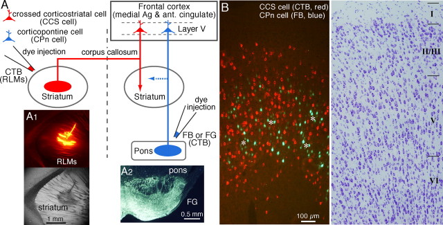
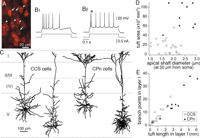
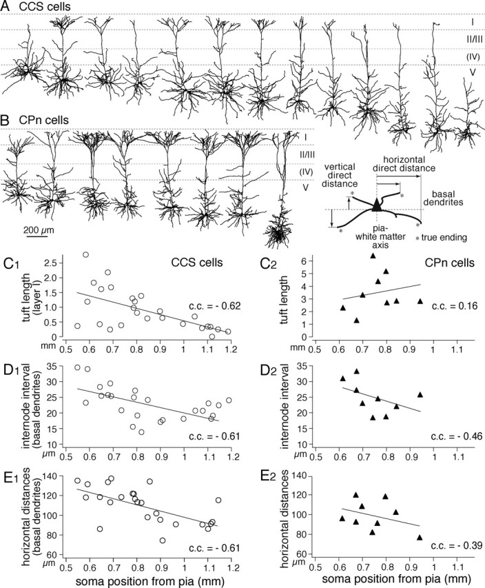
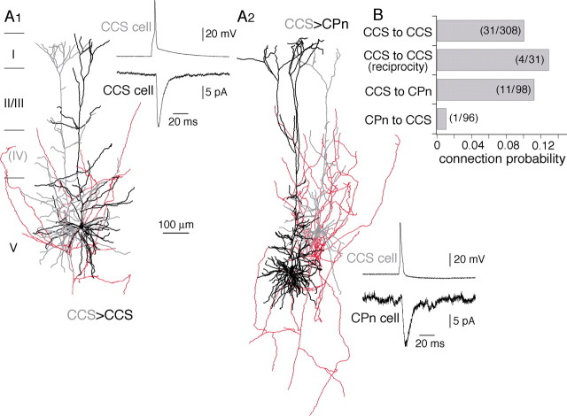
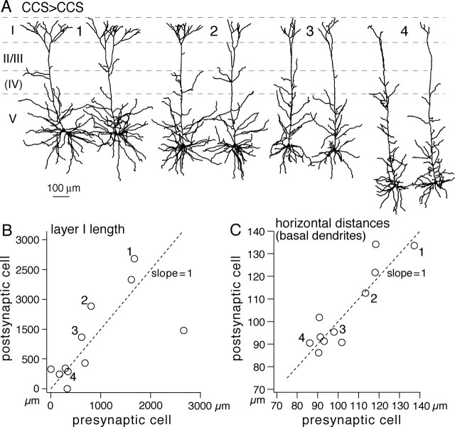
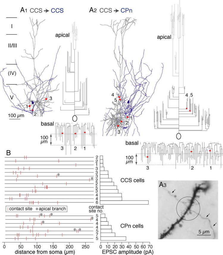
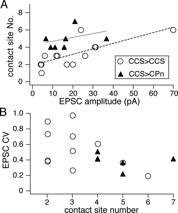
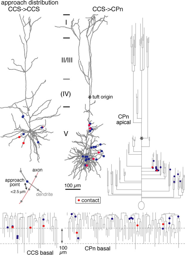
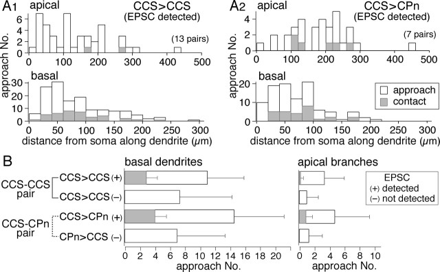
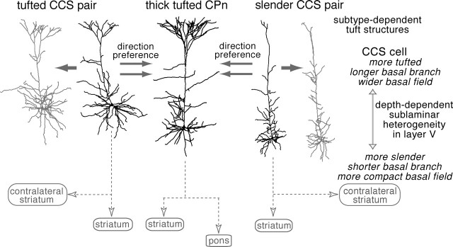
Corticostriatal pyramidal cells are heterogeneous in the frontal cortex. Here, we show that subpopulations of corticostriatal neurons in the rat frontal cortex are selectively connected with each other based on their subcortical targets. Using paired recordings of retrogradely labeled cells, we investigated the synaptic connectivity between two projection cell types: those projecting to the pons [corticopontine (CPn) cell], often with collaterals to the striatum, and those projecting to both sides of the striatum but not to the pons [crossed corticostriatal (CCS) cell]. The two types were morphologically differentiated in regard to their apical tufts. The dendritic morphologies of CCS cells were correlated with their somatic depth within the cortex. CCS cells had reciprocal synaptic connections with each other and also provided synaptic input to CPn cells. However, connections from CPn to CCS cells were rarely found, even in pairs showing CCS to CPn connectivity. Additionally, CCS cells preferentially innervated the basal dendrites of other CCS cells but made contacts onto both the basal and apical dendrites of CPn cells. The amplitude of synaptic responses was to some extent correlated with the contact site number. Ratios of the EPSC amplitude to the contact number tended to be larger in the CCS to CCS connection. Therefore, our data demonstrate that these two types of corticostriatal cells distinct in their dendritic morphologies show directional and domain-dependent preferences in their synaptic connectivity.


Figures

Figure 1.
Figure 1.
Two projection types of pyramidal cells in layer V of the frontal cortex. A, Schematic of method for identification of two projection neuron types by retrograde tracers. Alexa Fluor 555-conjugated CTB was injected in the contralateral striatum while Fast Blue (FB) or Fluorogold (FG) was injected into the pontine nuclei. For simultaneous recording from CCS and CPn cells, RLMs were injected in the contralateral striatum, and CTB was injected in the ipsilateral pons. Ag, Agranular; ant., anterior. A1, Injection site of RLMs in the striatum. Top, Epifluorescence view. Bottom, As in above, but using bright-field microscopy. The tracer was applied obliquely through the lateral ventricle after suctioning the overlying cortex to prevent injection into the cortex. A2, Injection site of Fluorogold in the pons including the ipsilateral pontine nuclei. B, Fluorescence imaging revealed two nonoverlapping populations of CCS and CPn cells. CPn cells were distributed in patchy regions (asterisks) where CCS cells were absent. Right, As in the left but in thionin staining.
Figure 2.
Figure 2.
Morphological characteristics of the two projection types of pyramidal cells. A, Simultaneous fluorescent labeling of CCS cells with RLMs (arrowheads) and CPn cells with CTB (arrows). B1, B2, Firing responses of CCS and CPn cells, respectively. Note the initial doublet firing (*) in a CPn cell. C, Dendritic reconstructions of two CCS and two CPn cells, showing the differences in dendritic patterns, especially in their apical tufts. D, Plot of the differences in the apical tuft area and shaft diameter between CCS (open circles) and CPn (filled triangles) cells. E, Differences in the dendritic length of layer I tufts and branch points in layer I between CCS and CPn cells.
Figure 3.
Figure 3.
Depth dependency of dendritic morphologies among CCS cells. A, B, Dendritic reconstructions of CCS cells (top) and CPn cells (bottom), arranged from most superficial to deeper somata in layer V. C–E, Depth profiles of dendritic lengths in layer I (C), mean internode intervals of basal dendrites (D), and horizontal direct distances of basal dendrite endings (E) (inset defines the parameters of endings). Note the depth dependency of these parameters in CCS cells. Lines are linear fits.
Figure 4.
Figure 4.
Synaptic connections among corticostriatal pyramidal cells. A1, Reconstructions of the dendrites of presynaptic (gray) and postsynaptic (black) CCS cells and the axon of the presynaptic CCS cell (red). Inset, A presynaptic action potential (top trace) generates a unitary postsynaptic current (bottom trace). A2, Reconstructions and postsynaptic current of a presynaptic CCS cell (gray) and postsynaptic CPn (black) cells. B, Connection probability between pyramidal cell subtypes. Note that very few connections were found from CPn to CCS cells.
Figure 5.
Figure 5.
Synaptically connected CCS cells show similar dendritic morphologies. A, Dendritic reconstructions of four connected CCS cell pairs (1–4). The left is presynaptic cells. In pair 4, the cells were bidirectionally connected. Spatially overlapped neurons in the pairs are separate to see individual morphologies. Note the comparable dendritic patterns between CCS cell pairs. B, Comparison of dendritic lengths in layer I between presynaptic and postsynaptic cells. C, Comparison of mean horizontal direct distances of endings (Fig. 3, inset). Note the presynaptic and postsynaptic dendritic similarity. Numbers (1–4) in B and C correspond to those of pairs in A.
Figure 6.
Figure 6.
Contact sites between presynaptic axons and postsynaptic dendrites. A, Examples of dendritic contact sites in a CCS to CCS connection (A1; 1–3) and a CCS to CPn connection (A2; 1–5). Dendrites are shown in gray, and axons are shown in blue. Contact sites (red circles) are shown on the dendrograms of the apical and basal dendrites. Apical tufts are shown in light gray. A3, Photograph of a contact site between axon and dendrite in a CCS to CCS pair. The presynaptic axon is marked by arrows. B, Plot of the distribution of contact sites along the dendrites of 13 CCS and 7 CPn cells (left) and the corresponding EPSC amplitude (right). Contacts on apical branches are marked by asterisks.
Figure 7.
Figure 7.
Relationship of contact site number and EPSC characteristics. A, Relationship of contact site numbers with the corresponding EPSC amplitudes. The correlation in CCS to CCS pairs (linear fitting line in red) was stronger than that in CCS to CPn pairs (linear fitting line in gray). B, Relationship of CV of EPSC amplitudes with the corresponding number of morphological contact sites, which were inversely correlated (c.c., −0.51; p < 0.05).
Figure 8.
Figure 8.
Distributions of approach points along reconstructed dendrites and dendrograms of example postsynaptic neurons (basal dendrites of a CCS cell, and basal and apical dendrites of a CPn cell). Contact sites are shown in red, and noncontact approach points are shown in blue. Inset, Example of an approach point (blue circle) of an axon (red line) to a dendrite (gray line). Reconstructed axons and dendrites were composed of serial points (circles) with intervals shorter than 1.5 μm. Approach points were defined as dendritic sites where axons passed within 2.5 μm.
Figure 9.
Figure 9.
Frequency of axonal contacts onto nearby postsynaptic dendrites. A1, A2, Dendritic distance distributions of total approaches and contacts along dendrites from soma in connected CCS to CCS (A1; from 13 cells) and CCS to CPn (A2; from 7 cells) pairs; those at the top are in apical branches, and those at the bottom are in basal dendrites. The distribution patterns were similar between approaches and contacts in basal dendrites of connected pairs. Apical branch contacts were more frequent in connected CCS to CPn than in CCS to CCS pairs. B, Mean number of approach points and contact sites in CCS–CCS and CCS–CPn pairs. Data are shown separately for basal and apical branches. Pairs with unitary EPSCs (marked by +) always showed contact sites. Unconnected pairs had no contact sites and fewer approaches. Note that contact site ratios in basal dendrites of both types and apical dendrites of CPn cells were similar (0.2–0.3), but those in apical dendrites of CCS cells were much lower. Data are means ± SD.
Figure 10.
Figure 10.
Innervation hierarchy and morphological pairing in synaptic connections between CCS and CPn cells. CCS cells were connected with each other and also with CPn cells, but connections from CPn to CCS cells were rarely found. Innervated domains were both basal and apical branches in the pairs from CCS to CPn cells but mostly basal dendrites in those between CCS cells. CCS cells were often connected with other CCS cells with similar dendritic patterns. CCS cells were heterogeneous in apical tuft and basal structures, which were dependent on the somatic depth from pia.

References

    1. Albin RL, Young AB, Penney JB (1989). The functional anatomy of basal ganglia disorders. Trends Neurosci 12:366–375. - PubMed
    1. Alexander GE, Crutcher MD (1990). Functional architecture of basal ganglia circuits: neural substrates of parallel processing. Trends Neurosci 13:266–271. - PubMed
    1. Anderson J, Lampl I, Reichova I, Carandini M, Ferster D (2000). Stimulus dependence of two-state fluctuations of membrane potential in cat visual cortex. Nat Neurosci 3:617–621. - PubMed
    1. Bauswein E, Fromm C, Preuss A (1989). Corticostriatal cells in comparison with pyramidal tract neurons: contrasting properties in the behaving monkey. Brain Res 493:198–203. - PubMed
    1. Christophe E, Doerflinger N, Lavery DJ, Molnar Z, Charpak S, Audinat E (2005). Two populations of layer V pyramidal cells of the mouse neocortex: development and sensitivity to anesthetics. J Neurophysiol 94:3357–3367. - PubMed

Publication types