

Isolating the modulatory effect of expectation on pain transmission: a functional magnetic resonance imaging study
- PMID: 16624963
- PMCID: PMC6674009
- DOI: 10.1523/JNEUROSCI.4463-05.2006
Isolating the modulatory effect of expectation on pain transmission: a functional magnetic resonance imaging study
Abstract
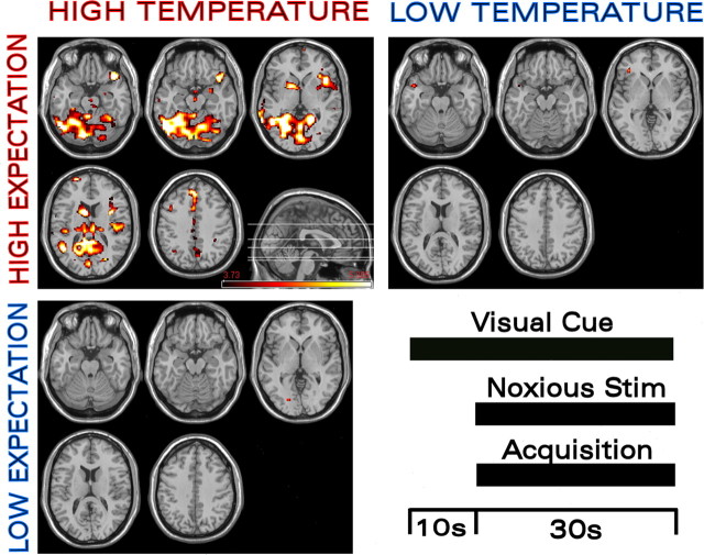
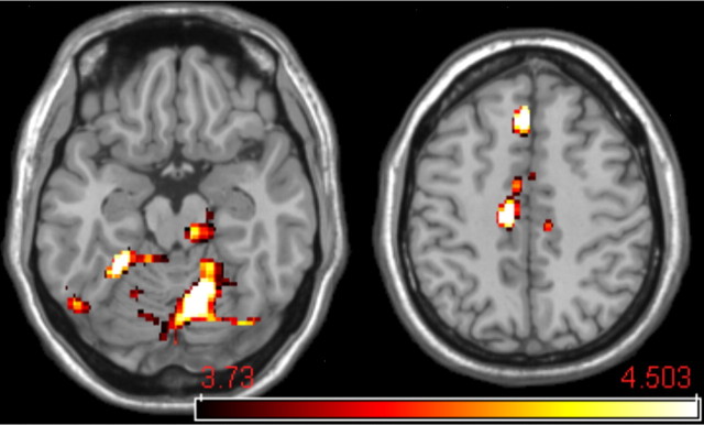
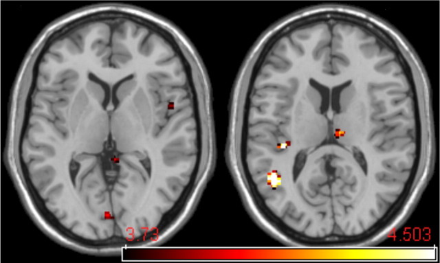
We use a novel balanced experimental design to specifically investigate brain mechanisms underlying the modulating effect of expected pain intensity on afferent nociceptive processing and pain perception. We used two visual cues, each conditioned to one of two noxious thermal stimuli [ approximately 48 degrees C (high) or 47 degrees C (low)]. The visual cues were presented just before and during application of the noxious thermal stimulus. Subjects reported significantly higher pain when the noxious stimulus was preceded by the high-intensity visual cue. To control for expectancy effects, for one-half of the runs, the noxious thermal stimuli were accompanied by the cue conditioned to the other stimulus. Comparing functional magnetic resonance imaging blood oxygenation level-dependent activations produced by the high and low thermal stimulus intensities presented with the high-intensity visual cue showed significant activations in nociceptive regions of the thalamus, second somatosensory cortex, and insular cortex. To isolate the effect of expectancy, we compared activations produced by the two visual cues presented with the high-intensity noxious thermal stimulus; this showed significant differences in the ipsilateral caudal anterior cingulate cortex, the head of the caudate, cerebellum, and the contralateral nucleus cuneiformis (nCF). We propose that pain intensity expectancy modulates activations produced by noxious stimuli through a distinct modulatory network that converges with afferent nociceptive input in the nCF.
Figures
References
-
- Bandler R, Shipley MT (1994). Columnar organization in the midbrain periaqueductal gray: modules for emotional expression? Trends Neurosci 17:379–389. - PubMed
-
- Bantick SJ, Wise RG, Ploghaus A, Clare S, Smith SM, Tracey I (2002). Imaging how attention modulates pain in humans using functional MRI. Brain 125:310–319. - PubMed
-
- Benedetti F, Amanzio M, Casadio C, Oliaro A, Maggi G (1997). Blockade of nocebo hyperalgesia by the cholecystokinin antagonist proglumide. Pain 71:135–140. - PubMed
-
- Bingel U, Lorenz J, Schoell E, Weiller C, Buchel C (2006). Mechanisms of placebo analgesia: rACC recruitment of a subcortical antinociceptive network. Pain 120:8–15. - PubMed
-
- Bushnell MC, Apkarian AV (2005). Representation of pain in the brain. In: Wall and Melzack’s textbook of pain, Chap 6 (McMahon SB, Koltzenburg M, eds) pp. 107–124. New York: Elsevier, Churchill Livingstone.
Publication types
MeSH terms
Grants and funding
LinkOut - more resources
Full Text Sources
Medical