. 2006 Jun 2;359(2):486-95.
doi: 10.1016/j.jmb.2006.03.039. Epub 2006 Mar 30.

Structural bioinformatics prediction of membrane-binding proteins

Affiliations

Structural bioinformatics prediction of membrane-binding proteins

Nitin Bhardwaj et al. J Mol Biol. .

Abstract

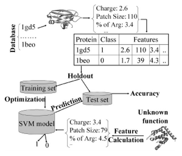
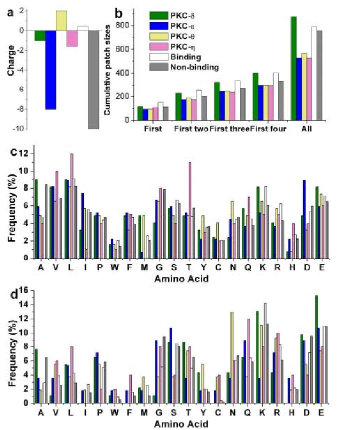
Membrane-binding peripheral proteins play important roles in many biological processes, including cell signaling and membrane trafficking. Unlike integral membrane proteins, these proteins bind the membrane mostly in a reversible manner. Since peripheral proteins do not have canonical transmembrane segments, it is difficult to identify them from their amino acid sequences. As a first step toward genome-scale identification of membrane-binding peripheral proteins, we built a kernel-based machine learning protocol. Key features of known membrane-binding proteins, including electrostatic properties and amino acid composition, were calculated from their amino acid sequences and tertiary structures, which were then incorporated into the support vector machine to perform the classification. A data set of 40 membrane-binding proteins and 230 non-membrane-binding proteins was used to construct and validate the protocol. Cross-validation and holdout evaluation of the protocol showed that the accuracy of the prediction reached up to 93.7% and 91.6%, respectively. The protocol was applied to the prediction of membrane-binding properties of four C2 domains from novel protein kinases C. Although these C2 domains have 50% sequence identity, only one of them was predicted to bind the membrane, which was verified experimentally with surface plasmon resonance analysis. These results suggest that our protocol can be used for predicting membrane-binding properties of a wide variety of modular domains and may be further extended to genome-scale identification of membrane-binding peripheral proteins.


Figures

Fig 1
Fig 1
Overall strategy for constructing the protocol for identification of membrane-binding proteins. For each protein in the database (composed of negative and positive cases), features are generated. The dataset is then divided into two parts for holdout evaluation (the complementary cross-validation evaluation is not shown here). The SVM is optimized for best separation on the training set, which is then tested using the testing set. For an unclassified protein, similar features are generated and fed into the SVM for classification (1 or 0).
Fig 2
Fig 2
Box and whisker plots showing the distribution of net charge (a) and cumulative positive patch sizes (b) for lipid-binding (clear) and non-binding proteins (shaded). Fisher's score for individual features are also shown.
Fig 3
Fig 3
Relative orientation of cationic patches with respect to membrane binding surfaces. For cytosolic phospholipase A2 C2 domain (A) and PKCα C2 domain (B), membrane binding surfaces were experimentally determined. For β-spectrin PH domain (C), CALM-ANTH domain (D), and epsin 1-ENTH domain (E), membrane binding surfaces have been proposed based on biophysical and mutation studies. Proteins are shown in cartoon representation and cationic patches are displayed in ‘surface’ representation. RGB scale is used to color the patches with blue, cyan, white, and pink representing the largest, the second largest, third largest, and the fourth largest patches, respectively. A solid line indicates the location of the lipid headgroup region in the lipid bilayer. Figure are made with VMD .
Fig 4
Fig 4
Histograms showing the overall (a) and surface (b) amino acid composition of binding (clear) and non-binding (shaded) proteins. Corresponding compositions for all proteins in the Astral-40 database are also indicated (black bars). The solid line plots the ratio of frequency in binding proteins to that in non-binding proteins. The ratio above 1.0 (see the right Y axis) indicates a higher propensity to be present in membrane-binding proteins.
Fig 5
Fig 5
Distribution of various features for the C2 domains of PKCδ, ε, η and θ. The average value of corresponding features from the known binding and non-binding groups are shown for a direct comparison of the former with the any of the latter two cases.
Fig 6
Fig 6
SPR Sensorgrams four PKC C2 domains. SPR sensorgrams were obtained by monitoring resonance unit (RU) changes after injecting each C2 domain (25 nM for PKCθ-C2 and 5 μM for other C2 domains) to the sensor chip coated with POPC/POPS/PtdIns(4,5)P2 (65:30:5) vesicles at 30 μl/min. The control surface was coated with 100% POPC vesicles. 10 mM HEPES buffer, pH 7.4, with 0.16 M NaCl was used for these measurements.

References

    1. Cho W, Stahelin RV. Membrane-protein interactions in cell signaling and membrane trafficking. Annu Rev Biophys Biomol Struct. 2005;34:119–51. - PubMed
    1. Teruel MN, Meyer T. Translocation and reversible localization of signaling proteins: a dynamic future for signal transduction. Cell. 2000;103:181–4. - PubMed
    1. Hurley JH, Meyer T. Subcellular targeting by membrane lipids. Curr Opin Cell Biol. 2001;13:146–52. - PubMed
    1. DiNitto JP, Cronin TC, Lambright DG. Membrane recognition and targeting by lipid-binding domains. Sci STKE. 2003;2003:re16. - PubMed
    1. Cho W. Membrane targeting by C1 and C2 domains. J Biol Chem. 2001;276:32407–10. - PubMed

Publication types

Substances

LinkOut - more resources