Review
. 2006 May 29;361(1469):821-34.
doi: 10.1098/rstb.2006.1831.

Ovulation, pregnancy, placentation and husbandry in the African elephant (Loxodonta africana)

Affiliations
Review

Ovulation, pregnancy, placentation and husbandry in the African elephant (Loxodonta africana)

W R Allen. Philos Trans R Soc Lond B Biol Sci. .

Abstract

The African elephant reproduces so efficiently in the wild that overpopulation is now a serious problem in some game parks in Zimbabwe, Botswana and South Africa. The female reaches puberty between 10 and 12 years of age in the wild and, when in captivity, shows oestrous cycles of 14-15 weeks duration. She readily conceives a singleton in the wild yet her uterus has the capacity for twins. She shows a gestation length of 22 months and, in the wild, shows a population density and feed dependent intercalving interval of 4-8 years. The trophoblast erodes the lumenal epithelium of the endometrium and stimulates upgrowths of blood vessel-containing stromal villi, which develop eventually into the broad, tightly folded lamellae of the zonary, endotheliochorial placenta. Significant quantities of leaked maternal erythrocytes and ferric iron are phagocytosed by specialized trophoblast cells in the haemophagous zones at the lateral edges of the placental band. Although the placenta itself is endocrinologically inert, the foetal gonads, which enlarge greatly during the second half of pregnancy can synthesize 5alpha-dihydryoprogesterone and other 5alpha pregnane derivatives from cholesterol and pregnenolone. These products may synergize with progestagens secreted by the 2-8 large corpora lutea which are always present in the maternal ovaries throughout gestation to maintain the pregnancy state.


Figures

Figure 1
Figure 1
(a) The reproductive tract of a 4.5 month pregnant elephant viewed from the dorsal surface. The discrete conceptus bulge containing a foetus weighing 5.15 g is situated at the lateral divergence of the right uterine horn. (b) Photomicrograph of the endometrium of an adult non-pregnant female elephant showing the very close apposition of the lumenal epithelia to almost completely ablate the uterine lumen (Scale bar=90 μm). (c) Close-up view of a typical non-pregnant adult ovary extruded from its bursa to reveal the oedematous and velvety mucosal lining of the latter. (d) Four large pregnancy corpora lutea clustered on the ovary ipsilateral to the gravid uterine horn of an elephant at 6.5 months of gestation. (e) Unilateral conceptus bulge containing a foetus weighing 17 kg at 13.5 months of gestation.
Figure 2
Figure 2
(a) An opened conceptus sac that contained a 1.6 g foetus (four months gestation; Craig 1984). Note the pressurized bulging of the pale, tough endometrium with the attached embryonic membranes. The pale outline of the progenitor placental band is just visible (asterisk), in the middle of which primitive embryonic and placental blood vessels can be seen. (b) A conceptus bulge opened to reveal its 5.15 g foetus at 4.5 months of gestation. The separate allantoic locules can be seen and the transversely sectioned developing placental band is visible at the top of the conceptus (asterisk). (c) The gravid horn at 6.6 months of gestation opened to reveal the ovate conceptus now firmly attached to the endometrium by the thin fibrous-looking maternal placental hilus (asterisk) running in the centre along the length of the thickened and reddish-coloured placental band. Note the khaki-coloured homogenous luteal tissue of the two bisected corpora lutea in the ipsilateral ovary. (d) Close up view of (c) showing the maternal placental hilus being severed from the endometrium to allow the conceptus to roll out of the uterus. Note the relative narrowness of the maternal hilus and the great concentration of endometrial blood vessels feeding into it (asterisk). (e) The endometrial surface of one uterine horn in a very aged (more than 60 years) non-pregnant female, showing five concentric circumferential lines indicating the sites of attachment of the maternal placental hilus in previous pregnancies.
Figure 3
Figure 3
(a) High power photomicrograph of the trophoblast–endometrium interface in the every early stages of gestation (estimated 15–40 days, no embryo visible). A blunt projection of trophoblast cells (T) is forcing its way beneath the more darkly stained lumenal epithelial cells (E) and lifting them off the basement membrane (Scale bar=90 μm). (b) The endometrium–trophoblast interface in a second elephant in the very early stages of gestation (estimated 15–40 days, no embryo visible) showing the trophoblast cells (T) traversing down an endometrial gland by passing beneath and dehiscing the more darkly stained glandular epithelium (E). The basement membrane of the gland remains intact, thereby preventing the trophoblast cells from passing into the endometrial stroma (Scale bar=90 μm). (c) Low power photomicrograph of the developing placental band on a conceptus containing a 1.6 g foetus at four months gestation showing the line of elongating frond-like upgrowths of endometrial stroma, each covered by the single cell layer of trophoblast (Scale bar=30 μm). (d) Higher power photomicrograph of (c). At this early stage an appreciable amount of endometrial stroma (ES) separates the maternal capillaries from the trophoblast cells within each upgrowth, although far greater amounts of allantoic stroma (AS) containing the foetal blood vessels separate adjacent upgrowths (Scale bar=90 μm). (e) Low power photomicrograph at the base of the placental band on a conceptus carrying a 240 g foetus at 6.7 months of gestation. Note the development of the narrow attachment hilus and the continuing upward growth and stretching of the placental lamellae (Scale bar=30 μm). (f) Section of the placental band on a conceptus carrying a 1.9 kg foetus at 8.85 months gestation at the edge of the haemophagous zone. The specialist columnar trophoblast cells in this region are bathed continuously in extravasated maternal blood (Scale bar=90 μm).
Figure 4
Figure 4
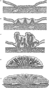
Diagrammatic representation of significant morphological changes that occur during the development of the zonary placental band on the elephant conceptus. (a) Replacement of the lumenal epithelium of the endometrium by trophoblast in the equatorial region of the conceptus. (b) Commencing upgrowth of trophoblast-covered stromal villi above the surface of the endometrium. (c) Elongation and branching of the trophoblast covered lamellae, each containing maternal capillaries surrounded by adequate amounts of stromal tissue. Foetal capillaries containing nucleated red blood cells are scattered throughout the allantoic mesoderm that separates adjacent lamellae. (d) The elongating lamellae at the lateral edge of the placental band lay over towards the endometrium to create the blind ending cleft in which the haemophagous zone will form. (e) In the mature placental band, the very elongated stromal lamellae become increasingly folded or pleated so as to maximize the available surface area of fetomaternal contact for placental exchange. Leakage of maternal blood into the lateral clefts creates the haemophagous zones in which the morphologically differentiated trophoblast cells imbibe maternal blood components.

References

    1. Adams C.E. Embryo transfer in rodents. In: Adams C.E, editor. Mammalian egg transfer. CRC Press; Boca Raton, FL: 1982. pp. 22–23.
    1. Allen W.R, Mathias S.S, Wooding F.B.P, Skidmore J.A, van Aarde R.J. Placentation in the African elephant, Loxodonta africana. I. Endocrinological aspects. Reproduction. 2002a;60:105–116. - PubMed
    1. Allen W.R, Wilsher S, Stewart F, Ousey J, Fowden A. The influence of maternal size on placental, fetal and postnatal growth in the horse. II Endocrinology of pregnancy. J. Endocrinol. 2002b;172:237–246. 10.1677/joe.0.1720237 - DOI - PubMed
    1. Allen W.R, Mathias S, Wooding F.B.P, van Aarde R.J. Placentation in the African elephant (Loxodonta africana). II. Morphological changes in the uterus and placenta throughout gestation. Placenta. 2003;24:598–617. 10.1016/S0143-4004(03)00102-4 - DOI - PubMed
    1. Allen W.R, Stout S, Ford M. Placentation in the African elephant, Loxodonta africana. IV. Growth and organisation of the fetal gonads. Reproduction. 2005;130:713–720. 10.1530/rep.1.00696 - DOI - PubMed

Publication types

LinkOut - more resources