. 2006 May 2;103(18):7100-5.
doi: 10.1073/pnas.0602184103. Epub 2006 Apr 20.

The primary function of RNA binding by the influenza A virus NS1 protein in infected cells: Inhibiting the 2'-5' oligo (A) synthetase/RNase L pathway

Affiliations

The primary function of RNA binding by the influenza A virus NS1 protein in infected cells: Inhibiting the 2'-5' oligo (A) synthetase/RNase L pathway

Ji-Young Min et al. Proc Natl Acad Sci U S A. .

Abstract

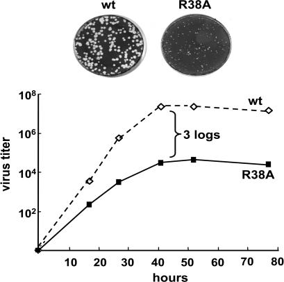
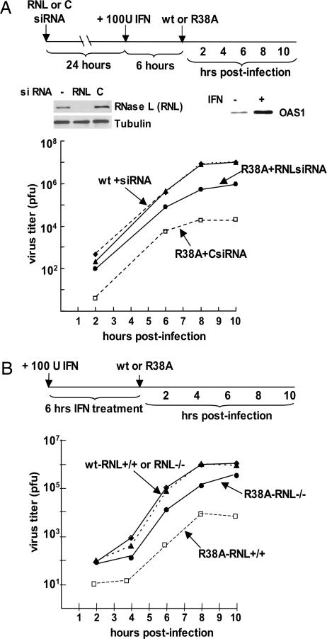
The NS1 protein of influenza A virus (NS1A protein) is a multifunctional protein that counters cellular antiviral activities and is a virulence factor. Its N-terminal RNA-binding domain binds dsRNA. The only amino acid absolutely required for dsRNA binding is the R at position 38. To identify the role of this dsRNA-binding activity during influenza A virus infection, we generated a recombinant influenza A/Udorn/72 virus expressing an NS1A protein containing an RNA-binding domain in which R38 is mutated to A. This R38A mutant virus is highly attenuated, and the mutant NS1A protein, like the WT protein, is localized in the nucleus. Using the R38A mutant virus, we establish that dsRNA binding by the NS1A protein does not inhibit production of IFN-beta mRNA. Rather, we demonstrate that the primary role of this dsRNA-binding activity is to protect the virus against the antiviral state induced by IFN-beta. Pretreatment of A549 cells with IFN-beta for 6 h did not inhibit replication of WT Udorn virus, whereas replication of R38A mutant virus was inhibited 1,000-fold. Using both RNA interference in A549 cells and mouse knockout cells, we show that this enhanced sensitivity to IFN-beta-induced antiviral activity is due predominantly to the activation of RNase L. Because activation of RNase L is totally dependent on dsRNA activation of 2'-5' oligo (A) synthetase (OAS), it is likely that the primary role of dsRNA binding by the NS1A protein in virus-infected cells is to sequester dsRNA away from 2'-5' OAS.


Conflict of interest statement

Conflict of interest statement: No conflicts declared.

Figures

Fig. 1.
Fig. 1.
The R38A influenza A/Udorn/72 mutant virus is attenuated. (Upper) Size of the plaques of the WT and R38A mutant viruses on MDCK cells. (Lower) Growth curve of the WT and R38A mutant viruses after low moi of MDCK cells.
Fig. 2.
Fig. 2.
Intracellular localization of the NS1A protein encoded by WT and R38A mutant influenza A/Udorn/72 viruses (Upper) and by WT and R38A, K41A mutant influenza A/WSN/33 viruses (Lower).
Fig. 3.
Fig. 3.
The Udorn R38A mutant virus exhibits no detectable defect during single-cycle virus growth and does not induce higher levels of IFN-β mRNA. (A) Viral protein synthesis after infecting MDCK cells with either WT or R38A Udorn virus at an moi of 5. Infected cells were labeled with [35S]methionine and [35S]cysteine at the indicated times after infection, and the radiolabeled proteins were analyzed by SDS/PAGE. The mobility of the R38A mutant NS1A protein is reduced relative to that of the WT NS1A protein. As a consequence, the R38A NS1A protein and the M1 protein have similar, but not identical, mobilities. These two proteins can be resolved by electrophoresis on higher concentration gels, which reveals that the relative amounts of M1 and NS1 are the same in WT and R38A virus-infected cells. (B) Growth curve of the WT and R38A mutant viruses after infecting MDCK cells at an moi of 5. (C) The relative amount of IFN-β mRNA produced during single-cycle growth in MDCK cells of WT, R38A mutant, and CPSF mutant viruses. At the times after infection indicated, cells were collected, and the total RNA was extracted. The amount of IFN-β mRNA was determined by using quantitative RT-PCR.
Fig. 4.
Fig. 4.
The Udorn R38A mutant virus has enhanced sensitivity to the antiviral state induced by IFN-β. A549 cells were treated or mock-treated with 100 units/ml IFN-β for 6 h. The cells were then infected with WT or R38A mutant virus at an moi of 5, and virus production was assayed as described in Materials and Methods.
Fig. 5.
Fig. 5.
Activation of the 2′-5′ OAS/RNase L pathway is the primary cause of the enhanced sensitivity of the R38A mutant virus to IFN-β. (A) siRNA knockdown of RNase L. A549 cells were transfected with either RNase L (RNL)-specific siRNAs or a control siRNA for 24 h. The cells were then treated with 100 units/ml IFN-β for 6 h, followed by infection with WT or R38A virus at an moi of 5. Virus production was assayed as described in Materials and Methods. The depletion of RNase L was assayed by an immunoblot of extracts of cells collected after IFN-β treatment. The production of OAS resulting from IFN-β treatment was monitored by immunoblotting using antibody against the OAS1 isomer (12). (B) Mouse RNase L knockout cells. Mouse RNase L−/− and RNase L+/+ cells were treated with 100 units/ml IFN-β for 6 h. The cells were then infected with WT or R38A mutant virus at an moi of 5, and virus production was assayed as described in Materials and Methods.

References

    1. Wright P. F., Webster R. G. In: Fields Virology. 4th Ed. Knipe D. M., Howley P. M., editors. Philadelphia: Lippincott Williams & Wilkins; 2001. pp. 1533–1579.
    1. Reid A. H., Taubenberger J. K., Fanning T. G. Microbes Infect. 2001;3:81–87. - PubMed
    1. Horimoto T., Kawaoka Y. Nat. Rev. Microbiol. 2005;3:591–600. - PubMed
    1. Noah D. L., Krug R. M. In: Advances in Virus Research. Maramorsch K., Shatkin A. J., editors. Vol. 65. Amsterdam: Elsevier; 2005. pp. 121–145. - PubMed
    1. Garcia-Sastre A. Virology. 2001;279:375–384. - PubMed

Publication types

LinkOut - more resources