

Prospective validation of a current algorithm including bedside US performed by emergency physicians for patients with acute flank pain suspected for renal colic
- PMID: 16627832
- PMCID: PMC2564078
- DOI: 10.1136/emj.2005.028589
Prospective validation of a current algorithm including bedside US performed by emergency physicians for patients with acute flank pain suspected for renal colic
Abstract
Objective: The purpose of this study was to validate an algorithm recommended by current literature for the patients with acute flank pain and evaluate the validity of bedside ultrasonography (US) performed by emergency physicians (EP) as a part of this algorithm.
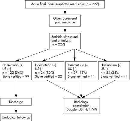
Materials and methods: This prospective validation study was carried out over a 5 month period in a tertiary care hospital adult emergency department (ED) with annual attendance of 55,000. Adult patients presenting to the ED with unilateral acute flank pain during the study period were enrolled into the study consecutively. Oral consent was obtained after the protocol was briefly explained to the patient and before the administration of analgesia. A protocol form was recorded for each patient enrolled into the study, and patients were followed up under the guidance of a previously designated algorithm in the ED. Data were analysed with SPSS software. The chi2 test was used to compare the dichotomised data of patients, diagnosed with and without stones, and to select the significant parameters to be used in the logistic regression.
Results: Of the 227 patients enrolled, 176 were proven to have urinary tract stones. There were 122 patients discharged from ED without further investigation except urinalysis and bedside US. Of these 122 directly discharged patients, 99 had a urinary stone, and the others did not have a life threatening disorder. Four of the 227 patients were admitted to the hospital. The remaining 51 patients did not have stones detected, and their pain subsided. Having a previous history of stones, radiation of pain to the groin, accompanying nausea, and detection of pelvicalyceal dilatation using bedside US performed by the EPs were found to be the most significant parameters in determining urinary stones in logistic regression analysis. Sensitivity and specificity of these parameters were: previous history of stones 59% and 66%, radiating pain to the groin 68% and 49%, nausea 71% and 51%, and detection of pelvicalyceal dilatation by bedside US 81% and 37%.
Conclusion: Bedside US performed by EPs could be used safely in the evaluation of patients with acute flank pain as a part of a clinical algorithm. Previous history of urinary stones, radiation of pain to the groin, accompanying nausea. and detection of pelvicalyceal dilatation are major parameters and symptoms of urinary stone disease, and could be used in the algorithms.
Conflict of interest statement
Competing interests: there are no competing interests
References
-
- Swadron S, Mandavia D P. Renal ultrasound. In: Ma OJ, Mateer JR, eds. Emergency ultrasound. New York: McGraw Hill Professional, 2002199
-
- Noble V E, Brown D F M. Renal ultrasound. Emerg Med Clin N Am 200422641–659. - PubMed
-
- Marston W A, Ahlquist R, Johnson G J.et al Misdiagnosis of ruptured abdominal aortic aneurysms. J Vasc Surg 19971617–22. - PubMed
-
- Pomper S R, Fiorillo M A, Anderson C W.et al Hematuria associated with ruptured abdominal aortic aneursym. Internat Surg 199580261–263. - PubMed
Publication types
MeSH terms
LinkOut - more resources
Full Text Sources
Other Literature Sources
Medical