. 2006 May;59(5):656-61.
doi: 10.1203/01.pdr.0000214958.80011.e1.

Breast milk lactoferrin regulates gene expression by binding bacterial DNA CpG motifs but not genomic DNA promoters in model intestinal cells

Affiliations

Breast milk lactoferrin regulates gene expression by binding bacterial DNA CpG motifs but not genomic DNA promoters in model intestinal cells

Peter Mulligan et al. Pediatr Res. 2006 May.

Abstract

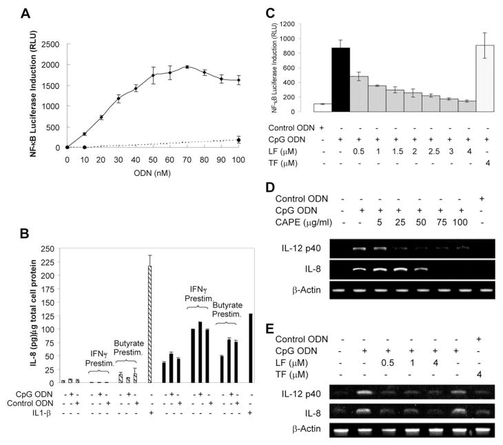
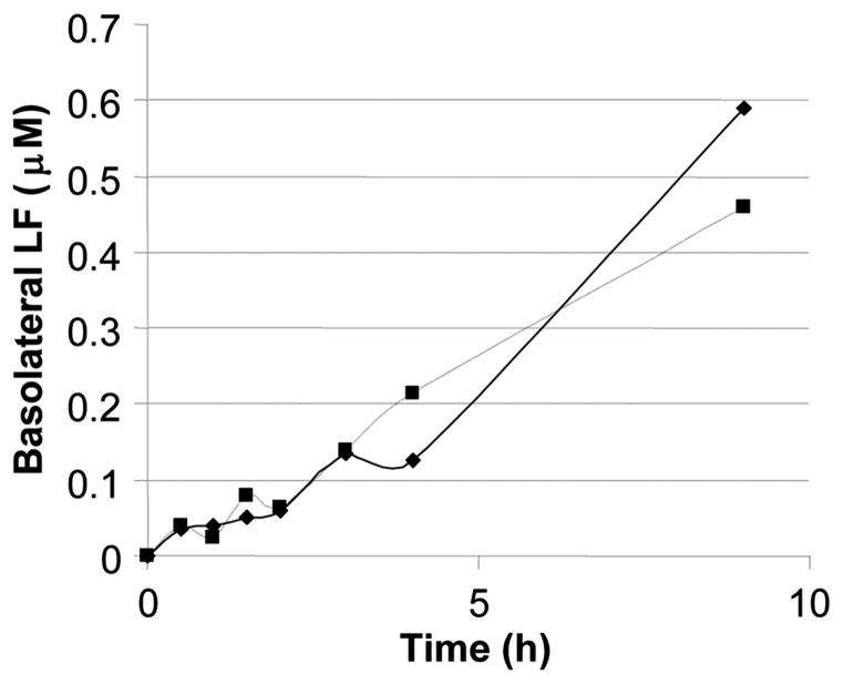
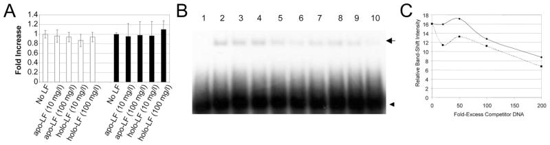
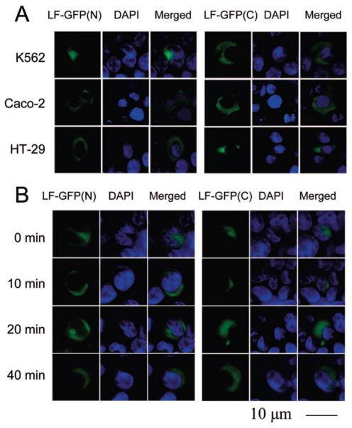
High-affinity binding of DNA by lactoferrin (LF) is an established phenomenon, but the biologic function of this interaction remains unclear. LF is an abundant breast milk protein (12.5-87.5 micromol/L) and is resistant to digestion in the infant gut. Regulation of gene expression by LF appears to be a major activity, particularly in modulating immune responses. We hypothesized that LF binding to DNA is a mechanism of gene regulation and aimed to identify the mechanism and physiologic sites of this activity. Our studies focused on two major biologic compartments of DNA: LF binding to proinflammatory bacterial DNA sequences (CpG motifs) in extracellular compartments and LF binding to genomic DNA promoters in the nucleus. LF 0.5 mmol/L inhibited CpG motif-induced nuclear factor-kappaB (NF-kappaB) activation and interleukin (IL)-8 and IL-12 cytokine gene transcription in B cells. Intestinal epithelial cells were unresponsive to CpG motifs. However, significant LF transferred across M cell-like monolayers, specialized epithelial cells that transcytose intact macromolecules to underlying B-cell follicles in the intestine. LF did not activate gene expression by binding to putative response elements in epithelial and lymphoid cells. Nor did LF bind to putative response elements specifically in gel-shift assays. No nuclear localization of LF was detected in green fluorescent protein (GFP) tagging experiments. We conclude that breast milk LF regulates gene expression by binding CpG motifs extracellularly, with follicular B cells in the infant intestine a likely target.


Figures

Figure 1
Figure 1
LF inhibits NF-κB activation and transcription of NF-κB–regulated cytokines induced by CpG ODN in RPMI 8226 B cells. (A) NF-κB luciferase assays in transfected RPMI 8226 B cells demonstrated that CpG ODN (solid line) induces a linear activation of NF-κB in the range of 0.01 to 0.05 μmol/L CpG ODN. No activation was observed when cells were stimulated with control ODN (broken line). (B) No activation of IL-8 expression was observed in HT-29 (shaded bars) and Caco-2 cells (solid bars) stimulated with 1 mmol/L CpG ODN. IL-1β (2 ng/mL) was included as a positive control for IL-8 activation. (C) RPMI 8226 B cells transfected with an NF-κB luciferase reporter gene construct were preincubated with apo-LF for 1 h before stimulation with 30 nmol/L CpG ODN. Significant inhibition of NF-κB activation was observed at apo-LF concentrations ≥0.5 μmol/L, with complete inhibition at 4.0 μmol/L. No inhibition was observed with 4.0 μmol/L apo-TF. RLU: Relative luciferase units. Mean ± SD of triplicate samples (AC). (D) Incubation of RPMI 8226 B cells with the NF-κB inhibitor CAPE inhibited CpG ODN-induced IL-8 and IL-12 p40 gene transcription, as determined by RT-PCR. (E) LF inhibited CpG motif–induced IL-8 and IL-12 p40 gene transcription in RPMI 8226 B cells. No inhibition was observed with 4.0 μmol/L apo-TF. The results shown are representative of three separate experiments.
Figure 2
Figure 2
Transcytosis of LF across an M cell–like monolayer. Immunoreactive LF was transcytosed across an M cell–like monolayer in a time-dependent manner. After 9 h, basolateral concentrations of LF in the range of 0.5 to 0.8 μmol/L were achieved when the apical LF concentration was 10 μmol/L. Equivalent results were observed for apo-LF (solid line) and holo-LF (broken line), suggesting that iron saturation had no significant effect. TEER values before and after LF transcytosis were consistently in the range of 700–850 Ωcm2.
Figure 3
Figure 3
LF does not regulate reporter gene transcription via LFRE binding in K562 or Caco-2 cells and binds putative LFREs nonspecifically. (A) K562 cells (open bars) and Caco-2 cells (solid bars) were transfected with LFRE reporter gene vectors and stimulated with holo- or apo-LF at 10 or 100 μg/mL for 48 h. Cell lysates were assayed for luciferase activity. No significant difference between unstimulated and stimulated cells was observed for either K562 cells or Caco-2 cells. Results shown are mean ± SD of three independent experiments. Luciferase values were normalized by expressing luciferase relative to Renilla as a fraction of unstimulated samples. Shown are results for LFRE1; similar results were obtained for LFRE2. (B) Competition-binding EMSAs compared binding of LFRE and nonspecific DNA to LF. Lane 1 is free 32P-LFRE, lane 2 32P-LFRE preincubated with 120 ng apo-LF, lanes 3– 6 contain 120 ng apo-LF and 20-, 50,- 100-, and 200-fold molar excess of unlabeled LFRE, respectively; lanes 7–10 contain 120 ng LF and 20, 50, 100, and 200-fold excess nonspecific unlabeled DNA, respectively. Position of the band shift is indicated by the arrow and unbound probe by the arrowhead. Results for LFRE1 are shown; similar results were obtained for LFRE2 and LFRE3. Results are representative of three separate experiments. (C) The intensity of each band in B was quantified by densitometry and plotted against fold excess of unlabeled LFRE (solid line) or nonspecific DNA (broken line).
Figure 4
Figure 4
LF does not translocate from the cytoplasm to the nucleus in K562, Caco-2, and HT-29 cells. (A) GFP-tagged LF was expressed in transiently transfected K562, Caco-2, and HT-29 cells and subcellular localization determined by fluorescence microscopy. LF was tagged on either the N-terminus [LF-GFP(N)] or the C-terminus [LF-GFP(C)]. Staining with DAPI revealed the position of the nucleus in both transfected and untransfected cells. Results are representative of three separate experiments. (B) Stimulation of transfected cells with 100 nmol/L LF for 20, 40, or 60 min had no effect on LF-GFP subcellular localization. Shown are results for K562 cells. Similar results were observed for Caco-2 and HT-29 cells. Experiments were repeated three times.

References

    1. Querinjean P, Masson PL, Heremans JF. Molecular weight, single-chain structure and amino acid composition of human lactoferrin. Eur J Biochem. 1971;20:420–425. - PubMed
    1. Brock J. Lactoferrin: a multifunctional immunoregulatory protein? Immunol Today. 1995;16:417–419. - PubMed
    1. Lonnerdal B, Adkins Y. Developmental changes in breast milk protein composition during lactation. In: Sanderson IR, Walker WA, editors. Development of the Gastrointestinal Tract. BC Decker Inc; Hamilton: 2000. pp. 227–244.
    1. Broxmeyer HE, Smithyman A, Eger RR, Meyers PA, de Sousa M. Identification of lactoferrin as the granulocyte-derived inhibitor of colony-stimulating activity production. J Exp Med. 1978;148:1052–1067. - PMC - PubMed
    1. Brines RD, Brock JH. The effect of trypsin and chymotrypsin on the in vitro antimicrobial and iron-binding properties of lactoferrin in human milk and bovine colostrum. Unusual resistance of human apolactoferrin to proteolytic digestion. Biochim Biophys Acta. 1983;759:229–235. - PubMed

Publication types

MeSH terms