. 2006 May 17;25(10):2178-88.
doi: 10.1038/sj.emboj.7601102. Epub 2006 Apr 20.

A conserved pathway to activate BRCA1-dependent ubiquitylation at DNA damage sites

Affiliations

A conserved pathway to activate BRCA1-dependent ubiquitylation at DNA damage sites

Jolanta Polanowska et al. EMBO J. .

Abstract

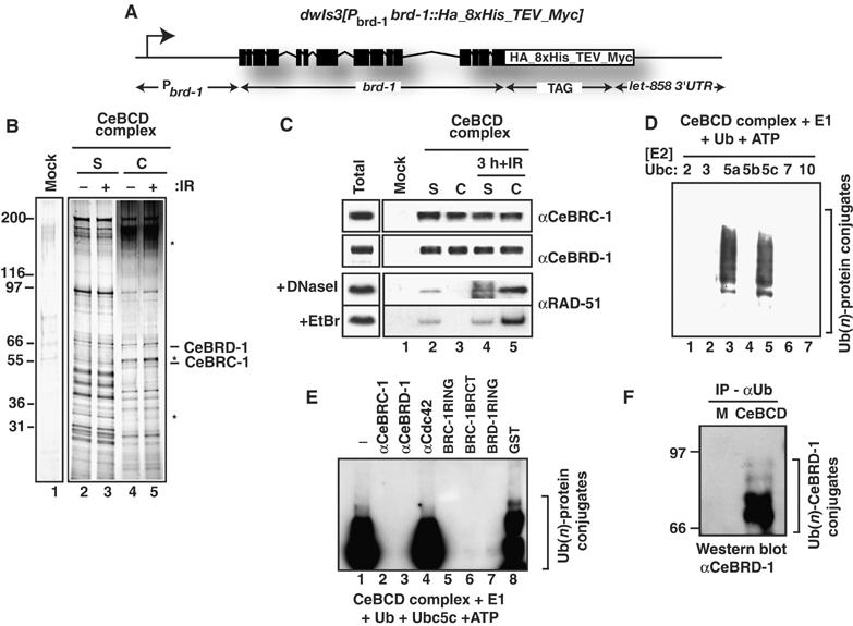
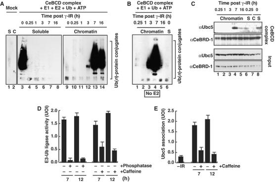
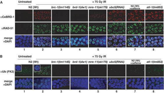
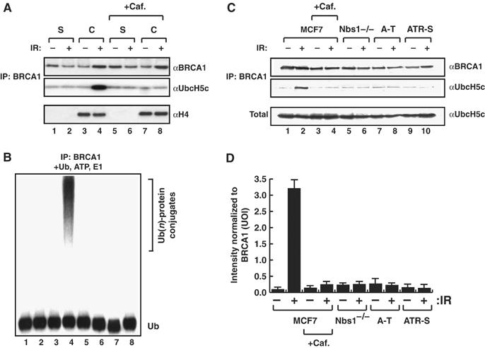
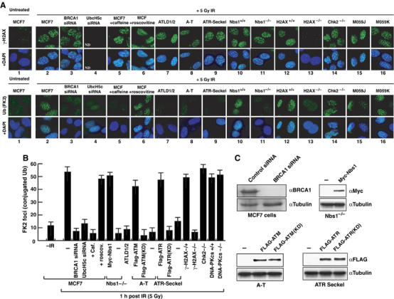
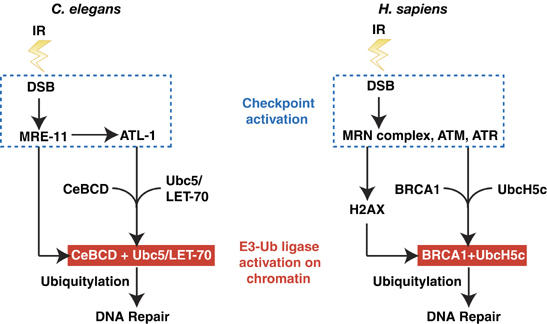
The BRCA1 tumour suppressor and its heterodimeric partner BARD1 constitute an E3-ubiquitin (Ub) ligase and function in DNA repair by unknown mechanisms. We show here that the Caenorhabditis elegans BRCA1/BARD1 (CeBCD) complex possesses an E3-Ub ligase responsible for ubiquitylation at DNA damage sites following ionizing radiation (IR). The DNA damage checkpoint promotes the association of the CeBCD complex with E2-Ub conjugating enzyme, Ubc5(LET-70), leading to the formation of an active E3-Ub ligase on chromatin following IR. Correspondingly, defects in Ubc5(let-70) or the DNA damage checkpoint genes atl-1 or mre-11 abolish CeBCD-dependent ubiquitylation in vivo. Extending these findings to human cells reveals a requirement for UbcH5c, the MRN complex, gamma-H2AX and a co-dependence for ATM and ATR kinases for BRCA1-dependent ubiquitylation at DNA damage sites. Furthermore, we demonstrate that the DNA damage checkpoint promotes the association between BRCA1 and UbcH5c to form an active E3-Ub ligase on chromatin after IR. These data reveal that BRCA1-dependent ubiquitylation is activated at sites of DNA repair by the checkpoint as part of a conserved DNA damage response.


Figures

Figure 1
Figure 1
CeBCD complex contains CeBRC-1, CeBRD-1 and RAD-51 and possess E3-Ub ligase activity. (A) The CeBRD-1 transgene used to purify the CeBCD complex. (B) Silver stain of the mock purification and CeBCD complexes following tandem immunoaffinity purification. Complexes shown have been normalized for CeBRC-1 and CeBRD-1. (S) Soluble and (C) chromatin bound, before and after IR-treatment. Asterisks mark bands enriched in the CeBCD complex (C) after IR-treatment. (C) Western blots of the mock purification and CeBCD complexes for CeBRC-1 and CeBRD-1. CeBCD complexes purified from extracts treated with DNaseI or ethidium bromide (EtBr) and Western blotted for RAD-51. (D) The CeBCD complex possesses E3-Ub ligase activity when incubated with recombinant E1 activating enzyme, ATP, Ub and recombinant UbcH5a or UbcH5c, but not with recombinant UbcH2, 3, 7 or 10. Poly-Ub conjugates were resolved on 10% PAGE gels and subjected to western blotting with anti-Ub antibodies. (E) E3-Ub ligase activity of the CeBCD complex was performed as in (D) following incubation with antibodies to CeBRC-1, CeBRD-1 or Cdc42 (lanes 2–4) or GST-fusions of the CeBRC-1-RING, CeBRC-1-BRCT, CeBRD-1-RING or GST alone (lanes 5–8). Poly-Ub conjugates were resolved on 10% PAGE gels and subjected to Western blotting with anti-Ub antibodies. (F) Western blot with αCeBRD-1 antibodies of mock (M) and CeBCD complexes following immunoprecipitation with αUb antibodies to enrich for ubiquitylated proteins.
Figure 2
Figure 2
CeBCD E3-Ub ligase is activated on chromatin in response to IR through a checkpoint-dependent interaction with Ubc5(LET-70). (A) E3-Ub ligase activity of CeBCD complexes purified from (S) Soluble and (C) chromatin bound extracts at the indicated times after IR. Assays were performed as in Figure 1D. (B) E3-Ub ligase activity of chromatin bound CeBCD complexes without addition of recombinant Ubc5 (E2) as a cofactor. (C) CeBCD complexes normalized for CeBRD-1 and Western blotted for Ubc5(LET-70) and CeBRD-1. (D) E3-Ub ligase activity of chromatin bound CeBCD complexes normalized with respect to CeBRC-1 and CeBRD-1, without addition of recombinant Ubc5 as a cofactor, 7 and 12 h after 75 Gy IR, without and with alkaline phosphatase or caffeine treatment. (E) Ubc5(LET-70) associated with chromatin bound CeBCD complexes normalized with respect to CeBRC-1 and CeBRD-1, 7 and 12 h after IR, without and with caffeine treatment. UOI, units of intensity. The averages (columns) and standard error of mean (s.e.m.: bars) of three independent experiments are presented. E3-Ub ligase activity assays were performed as in Figure 1D.
Figure 3
Figure 3
Conjugated Ub focus formation after IR is abolished in Cebrc-1, Cebrd-1, mre-11, atl-1 and Ubc5/let-70 mutants and after caffeine treatment. Representative images of fixed mitotic nuclei at the distal end of the germline stained for (A) CeBRD-1 and RAD-51, (B) conjugated-Ub (FK2) before (N2 wild type) and 3 h after 75 Gy IR-treatment for the indicated genotypes. brc-1(tm1145), brd-1(dw1), mre-11(ok179) and atl-1(tm853) are deletion mutants in C. elegans BRCA1, BARD1, MRE11 and ATR, respectively.
Figure 4
Figure 4
Human BRCA1 and UbcH5c associate in a checkpoint dependent manner to form an active E3-Ub ligase on chromatin. (A) UbcH5c and BRCA1 immunoblots of BRCA1 immunoprecipitates from MCF7 soluble (S) or chromatin bound (C) extracts before and 7 h after IR-treatment±caffeine treatment. The bottom panel is an immunoblot of the inputs with Histone H4 (chromatin marker) antibodies. (B) E3-Ub ligase assay performed on BRCA1 immunoprecipitates described in (A) in the presence of Ub, ATP and recombinant E1 activating enzyme (note: recombinant UbcH5c is not added to these reactions). Poly-Ub conjugates were resolved on 10% PAGE gels and subjected to western blotting with anti-Ub antibodies. The Ub-protein conjugates in lane 4 likely reflect autoubiquitylation of BRCA1. (C) UbcH5c and BRCA1 immunoblots of BRCA1 immunoprecipitates from MCF7 whole-cell extracts before and 7 h after IR-treatment±caffeine treatment, and from Nbs1−/−, A-T and ATR-Seckel whole-cell extracts before and 7 h after IR-treatment. Total is UbcH5c immunoblot of whole-cell extracts for each time point and genotype. (D) Quantification of UbcH5c association in BRCA1 immunoprecipitates shown in (C) normalized with respect to BRCA1. The averages (columns) and s.e.m. (bars) of two independent experiments are shown.
Figure 5
Figure 5
Genetic requirements for conjugated Ub focus formation after IR in mammalian cells. (A) Representative images of γ-H2AX, conjugated-Ub (FK2)±DAPI staining of DNA 1 h after 5 Gy of IR-treatment in cells of the indicated genotypes. ATLD1/2 cells are defective for Mre11; A-T cells are defective for Atm; ATR-Seckel cells carry a hypomorphic mutation in Atr; M059J (DNA-PKcs−/−), M059K (DNA-PKcs+/+). MCF7 cells were treated with 5 mM caffeine or 10 μM roscovitine for 6 h prior to IR-treatment. (B) Quantification of conjugated-Ub (FK2) focus formation 1 h post-treatment with 5 Gy of IR in cells of the indicated genotype. NBS1−/−, A-T and ATR-Seckel cells were complemented with the indicated plasmids: KD corresponds to kinase dead versions of ATM and ATR. The number of conjugated-Ub (FK2) foci was counted in at least 25 cells of each genotype. The averages (columns) and s.e.m. (bars) of three independent experiments are presented. (C) BRCA1 and Tubulin western blots of MCF7 extracts following control or BRCA1 siRNA. Western blots for Myc (Nbs1), Flag (ATM, ATM(KD), ATR, ATR(KD) and tubulin following correction of NBS1−/−, A-T and ATR-Seckel cell lines with the indicated plasmids. KD=kinase dead.
Figure 6
Figure 6
A conserved DNA damage-signalling pathway required for activation of BRCA1-dependent ubiquitylation during DNA repair. Following IR-induced damage, checkpoint activation in C. elegans and human cells promotes the association of CeBCD/BRCA1 with Ubc5/UbcH5c, respectively, and results in E3-Ub ligase activation. Once active, the E3-Ub ligase is responsible for ubiquitylation events at DNA damage sites. The sensitivity of Cebrc-1/BRCA1 mutants to DNA damaging agents suggest that their role in ubiquitylation at DNA damage sites may be required for efficient DNA repair.

References

    1. Alpi A, Pasierbek P, Gartner A, Loidl J (2003) Genetic and cytological characterization of the recombination protein RAD-51 in Caenorhabditis elegans. Chromosoma 112: 6–16 - PubMed
    1. Boulton SJ, Gartner A, Reboul J, Vaglio P, Dyson N, Hill DE, Vidal M (2002) Combined functional genomic maps of the C. elegans DNA damage response. Science 295: 127–131 - PubMed
    1. Boulton SJ, Martin JS, Polanowska J, Hill DE, Gartner A, Vidal M (2004) BRCA1/BARD1 orthologs required for DNA repair in Caenorhabditis elegans. Curr Biol 14: 33–39 - PubMed
    1. Bradbury JM, Jackson SP (2003) ATM and ATR. Curr Biol 13: R468. - PubMed
    1. Brenner S (1974) The genetics of Caenorhabditis elegans. Genetics 77: 71–94 - PMC - PubMed

Publication types

MeSH terms