. 2006 May 17;25(10):2119-30.
doi: 10.1038/sj.emboj.7601101. Epub 2006 Apr 20.

The FUSE/FBP/FIR/TFIIH system is a molecular machine programming a pulse of c-myc expression

Affiliations

The FUSE/FBP/FIR/TFIIH system is a molecular machine programming a pulse of c-myc expression

Juhong Liu et al. EMBO J. .

Abstract

FarUpStream Element (FUSE) Binding Protein (FBP) binds the human c-myc FUSE in vitro only in single-stranded or supercoiled DNA. Because transcriptionally generated torsion melts FUSE in vitro even in linear DNA, and FBP/FBP Interacting Repressor (FIR) regulates transcription through TFIIH, these components have been speculated to be the mechanosensor (FUSE) and effectors (FBP/FIR) of a real-time mechanism controlling c-myc transcription. To ascertain whether the FUSE/FBP/FIR system operates according to this hypothesis in vivo, the flux of activators, repressors and chromatin remodeling complexes on the c-myc promoter was monitored throughout the serum-induced pulse of transcription. After transcription was switched on by conventional factors and chromatin regulators, FBP and FIR were recruited and established a dynamically remodeled loop with TFIIH at the P2 promoter. In XPB cells carrying mutant TFIIH, loop formation failed and the serum response was abnormal; RNAi depletion of FIR similarly disabled c-myc regulation. Engineering FUSE into episomal vectors predictably re-programmed metallothionein-promoter-driven reporter expression. The in vitro recruitment of FBP and FIR to dynamically stressed c-myc DNA paralleled the in vivo process.


Figures

Figure 1
Figure 1
DNA binding by FBP requires sustained superhelical stress, whereas transient stress is sufficient for FIR. (A) Schematic drawing of liner in vitro transcription template. Active transcription, even in linear templates, develops dynamic supercoils that convert duplex FUSE (box) into single-stranded DNA (oval) (Kouzine et al, 2004). (B) Negative torsional stress (supercoiling) was dynamically generated by T3 and T7 RNA polymerase transcription in vitro of linear templates in the presence of FBP (α-FBP IP panel) or FIR (α-FIR IP panel). Primer extension (PE, lanes 4–8) showed increased transcription with increasing (GTP). EDTA was added to stop transcription prior to fixation (lane 9) to test if ongoing transcription was required for FBP or FIR binding. Immunoprecipitated DNA was amplified (IP, lanes 1–9). Real-time PCR profile of the same DNA is shown on the right. (C) In vitro KMnO4 footprints of FBP and FIR binding to supercoiled or linear DNA. Hyper-reactive residues are marked with triangles; hyper-reactive residues resistant to linearization are marked with *. Recombinant GST, FBP, and/or FIR were incubated with supercoiled (lanes 1–4 and lanes 9–12) or linear (lanes 5–8) DNA-containing FUSE. After binding, all samples received MgSO4 (2 mM final) and XhoI was added in lanes 9–12 for 5 min before KMnO4 treatment.
Figure 2
Figure 2
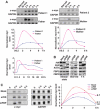
Complex transcription factor choreography precedes FBP and FIR recruitment to the c-myc promoter. Human Hs68 primary fibroblasts were cultured in DMEM without FBS for 5 days and then stimulated with 10% serum; cells were harvested for RNA or ChIP at the indicated times after adding serum. (A) RNase protection assay measuring c-myc levels. (B, C) Chromatin from starved and stimulated cells was immunoprecipitated with indicated antibodies. (D) Real-time PCR amplification of the same ChIP DNA samples shown in (C) (2 and 4 h). (E) Schematic representation of transcription factor binding sites and primer sets for PCR amplification.
Figure 3
Figure 3
Chromatin remodeling accompanies shutdown and reactivation of FUSE. (A) Southern blot of DNA from MNase-treated nuclei from serum-starved (lanes 1 and 2) or stimulated (lanes 3–8) primary fibroblasts reveals imprecise positioning of the FUSE nucleosome in activated cells. Nucleosomes are numbered as in Michelotti et al (1996). Nuclei were digested with MNase, and then cleaved with KpnI, separated electrophoretically and analyzed by Southern blot with a c-myc probe. (B) Southern blot showing nucleosome ladder around FUSE in IMR32 nuclei. (C) Fine-mapping of the FUSE-masking nucleosome. Mono-nucleosome-sized DNA from MNase-digested IMR32 nuclei was gel purified and subjected to LM–PCR. The 5′ (lanes 1 and 2) and 3′ (lanes 3 and 4) boundaries were defined by reverse and forward primer sets, respectively. No PCR product was obtained without adapter ligation (lane 5). (D) LM–PCR reveals imprecise repositioning of the nucleosome near FUSE in SW13 or U2OS cells expressing c-myc. (E) ChIP comparing FBP/FIR binding to FUSE in the presence (U2OS) or absence (SW13) of the BAF complex. (F) Transcription is necessary to hold FBP and FIR at FUSE. ChIP showing failure of FBP and FIR to bind FUSE in IMR32 cells, where c-myc is permanently silenced, although FBP and FIR are expressed at similar levels (Western panel).
Figure 4
Figure 4
TFIIH-mediated loop between FUSE and P2. (A) ChIP with indicated antibodies of HeLa chromatin. Amplicons were described in Figure 2E. (B) Mutation of the p89 (XPB) TFIIH subunit disrupts the loop. ChIP comparing log-phase XPB lymphoblasts (GM02252) and complemented cells. (C) An independent p89 (XPB) mutation also disrupts the loop. ChIP of c-myc-bound factors from log-phase primary fibroblasts from an XPB patient (GM13025) and parent (GM13028).
Figure 5
Figure 5
Looping between FUSE–FBP–FIR and TFIIH is essential for the normal response of c-myc to serum. (A) Independent RNase protection assays comparing the c-myc serum response of primary fibroblasts of XPB patients or their parents. c-myc mRNA levels were analyzed by ImageQuant and normalized to GAPDH (graph). c-myc level (normalized to γ-tubulin) was also monitored throughout a 24-h period by real-time RT–PCR (bottom). (B) Steady-state, serum-starved human Hs68 primary fibroblasts were transfected with siRNA to FIR or FBP. Starved cells were harvested before or after 4 h of serum repletion and subjected to Western blot. (C) RNase protection assay showing the serum response of c-myc in mock, siFBP, or siFIR transfected Hs68 cells (left). c-myc levels were analyzed by ImageQuant and normalized to GAPDH. Relative c-myc levels at 6 h post-serum induction are shown on the plot (right).
Figure 6
Figure 6
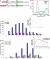
The FUSE–FBP–FIR system re-programs transcription from heterologous promoters. (A) Schematic drawing of reporters. (B) Raji cells carrying episomally encoded reporters under the control of identical, divergent metallothionein promoters (A, 3 kb separation) were induced with 90 μM Zn2+ for 4 h; fluorescence was analyzed by flow cytometry and a representative histogram of GFP fluorescence is shown. (C) Attenuation of FUSE activation after peak expression. Time course of median GFP fluorescence from cells with the 3 kb-separated promoters following Zn2+ induction. Ratio of +FUSE/−FUSE GFP fluorescence is also plotted (inset). (D) Median GFP fluorescence plot of either 3 or 4 kb separated reporters, with or without FUSE, at indicated time points. The ratio of the medians of GFP fluorescence with (+FUSE) or without (−FUSE) FUSE is also shown (inset).
Figure 7
Figure 7
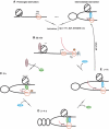
Proposed scheme for molecular servomechanism for regulation of c-myc transcription. (A) Without serum, c-myc transcription shuts off, FUSE becomes masked by a nucleosome, and only a trace of FIR remains. With prolonged starvation, FIR entirely disengages from FUSE. Even when shut off, a Pre-Promoter Escape Complex (PPC) remains paused within the promoter proximal region (P2). (B) The earliest factors binding to the promoter upon stimulation advance the PPC to a full Elongation Complex (EC), the FUSE-masking nucleosome is remodeled to expose FUSE. Negative supercoiling (−σ) generated by EC, combined with remodeling forces, initiate FUSE melting. (C) Melted FUSE recruits FBP, which drives transcription up to peak levels by looping with TFIIH. (D) FBP activity leads to negative supercoil accumulation within the topologically closed FBP–TFIIH loop. High −σ fully melts FUSE and conscripts FIR through protein–protein and protein–DNA contacts. (E) As FIR represses transcription, the dynamic stress at FUSE falls, ejecting FBP. The FIR–TFIIH connection allows only basal transcription; so the polymerase at a low rate dissociates from TFIIH to become an EC. Without sufficient activation, the superhelical stress eventually dissipates, and the c-myc promoter reverts back to the silent state in (A). For illustrative purposes, only the FUSE-masking nucleosome is shown.

Similar articles

Cited by

References

    1. Bazar L, Meighen D, Harris V, Duncan R, Levens D, Avigan M (1995) Targeted melting and binding of a DNA regulatory element by a transactivator of c-myc. J Biol Chem 270: 8241–8248 - PubMed
    1. Bentley DL, Groudine M (1986) A block to elongation is largely responsible for decreased transcription of c-myc in differentiated HL60 cells. Nature 321: 702–706 - PubMed
    1. Burgess-Beusse B, Farrell C, Gaszner M, Litt M, Mutskov V, Recillas-Targa F, Simpson M, West A, Felsenfeld G (2002) The insulation of genes from external enhancers and silencing chromatin. Proc Natl Acad Sci USA 99 (Suppl 4): 16433–16437 - PMC - PubMed
    1. Carey M, Smale ST (2000) Transcriptional Regulation in Eukaryotes: Concepts, Strategies, and Techniques. Cold Spring Harbor Laboratory Press: Cold Spring Harbor, NY
    1. Cawley S, Bekiranov S, Ng HH, Kapranov P, Sekinger EA, Kampa D, Piccolboni A, Sementchenko V, Cheng J, Williams AJ, Wheeler R, Wong B, Drenkow J, Yamanaka M, Patel S, Brubaker S, Tammana H, Helt G, Struhl K, Gingeras TR (2004) Unbiased mapping of transcription factor binding sites along human chromosomes 21 and 22 points to widespread regulation of noncoding RNAs. Cell 116: 499–509 - PubMed

Publication types

MeSH terms