. 2006 May 17;25(10):2155-66.
doi: 10.1038/sj.emboj.7601097. Epub 2006 Apr 20.

Interaction of Moloney murine leukemia virus matrix protein with IQGAP

Affiliations

Interaction of Moloney murine leukemia virus matrix protein with IQGAP

Juliana Leung et al. EMBO J. .

Abstract

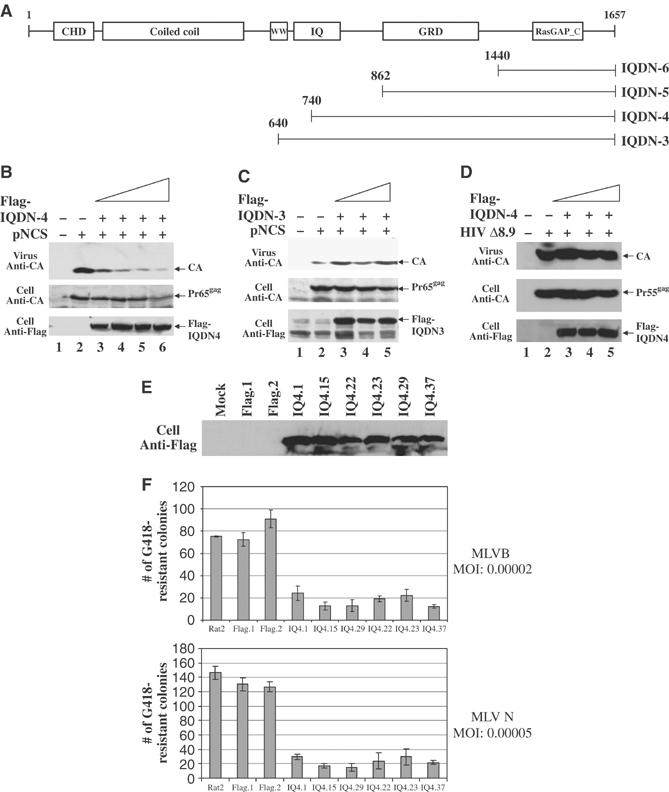
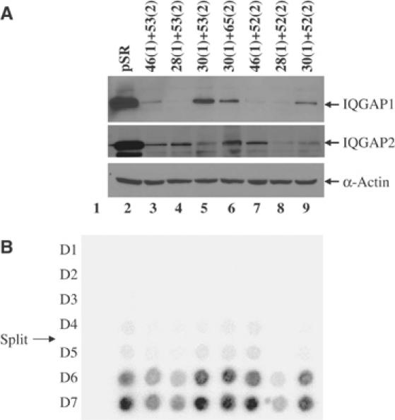
The matrix protein (MA) of the Moloney murine leukemia virus (M-MuLV) was found to interact with IQGAP1, a prominent regulator of the cytoskeleton. Mutational studies defined residues of MA critical for the interaction, and tests of viruses carrying MA mutations revealed a near-perfect correlation between binding and virus replication. The replication-defective mutants showed defects in both early and late stages of the life cycle. Four viable second-site revertant viruses were isolated from three different replication-defective parental mutants, and in all cases the interaction with IQGAP1 was restored by the suppressor mutations. The interaction of MA and IQGAP1 was readily detected in vitro and in vivo. Virus replication was potently inhibited by a C-terminal fragment of IQGAP1, and impaired by RNAi knockdown of IQGAP1 and 2. We suggest that the IQGAPs link the virus to the cytoskeleton for trafficking both into and out of the cell.


Figures

Figure 1
Figure 1
Construction and analysis of substitution mutants of M-MuLV MA. (A) Alanine-substitution mutations of MA. The complete sequence of MA is shown. Underlined resides were mutated to alanines in blocks of three, four, or five in the indicated mutants. (B) Assembly and release of mutant virions measured by RT assay. Spots indicate the amount of labeled nucleotide incorporated in an exogenous RT assay on a homopolymer template by virion-associated RT. Top row: 293T cells were transiently transfected by MA mutant proviral DNAs, and crude culture supernatants were harvested 48 h after transfection and analyzed by RT assay. Second row: virions were isolated from the medium by pelleting through a sucrose cushion, resuspended, and analyzed by RT assay. Mock, no viral DNA. pNCA, wild-type viral DNA. (C) Equal amounts of virus (normalized by RT assay of the crude harvests) were used to infect naïve Rat2 cells. Culture supernatants were harvested on successive days as indicated and virus yield was monitored by RT assay. Mock, no virus was applied. (D) Western blot analysis of CA-containing gag gene products in virions and lysates of transfected cells. The virus particles were prepared as described in Materials and methods. Virions and cells were lysed, and proteins were subjected to Western blot analysis, probing with anti-CA antisera. The cell lysates were normalized for transfection efficiency using β-galactosidase assay. The positions of Pr65gag and CA are indicated. (E) Southern blot detection of linear viral DNA synthesized in infected Rat2 cells. Supernatants from transfected 293T cells were used to acutely infect Rat2 cells. The virus preparations were normalized for equal RT activity. Low molecular weight DNAs were extracted by the Hirt method and the viral DNAs were detected by hybridization with a radiolabeled virus-specific DNA probe. The position of linear DNAs is marked. The levels of RT applied are shown in the lower panel.
Figure 2
Figure 2
DNA sequences, interaction phenotypes, and replication of MA revertants. (A) The amino-acid sequences of the MA proteins encoded by the wild-type virus, the parental mutants, and the revertant viral genomes. (B) Interaction of MA mutants and revertants with IQGAP1 in the yeast two-hybrid system. Signal strengths are as indicated in Table I. (C) The replication of wild-type, mutant, and revertant viruses in infected Rat2-2 cells as judged by RT assay of culture supernatants collected on the indicated days postinfection.
Figure 3
Figure 3
MA binding domain of IQGAP1 maps to the amino acids 1500–1657 (A) Deletion analysis. A schematic of the full-length IQGAP1 protein is shown at the top. The C-terminal region is expanded, and bars represent the portions expressed as Gal4AD fusions in yeast. Numbers refer to amino-acid residues of IQGAP1. Entries indicate level of β-galactosidase activity in yeast two-hybrid interaction assay, as given in Table I. (B) Alanine-scanning mutagenesis of selected regions of IQGAP1. Expanded views of amino acids 1440–1529 and 1642–1657 are shown. The position of the block of changes to alanine in each mutant is indicated. Yeast colonies expressing the indicated mutant fusion proteins were lifted and stained for β-galactosidase activity and scored as before.
Figure 4
Figure 4
(A) Interaction of MA with IQGAP1 in vitro. Wild-type MA, MA mutants (T4, T5, SMA6, and T11), and MA revertants (T4Rev, T1 Rev, T6Rev1, and T6Rev2) were expressed in bacteria as fusions with the MBP, bound to amylose–sepharose beads, and used as an affinity matrix for the binding of IQGAP1 expressed in bacteria as a fusion to (GST-IQGAP1; aa 1440–1657) or (GST-IQGAP1 Y1627H; aa 1440–1657). The beads were washed and the bound proteins were analyzed by SDS–PAGE and Western blot with anti-MA antisera and GST antibodies. (B) Pr65gag and IQGAP1 associate in vivo. 293T cells were cotransfected with either wild-type proviral DNA (pNCS) or the T4 mutant (pNCS-T4), and with either Flag-tagged full-length IQGAP1 or the C-terminal fragment Flag-IQDN4. Lysates were prepared, the Gag proteins were immunoprecipitated with anti-CA antiserum, and the associated proteins were analyzed by SDS–PAGE and Western blot with anti-FLAG antibodies and anti-CA antisera. As a negative control, samples were also immunoprecipiated with anti-HA antibodies. A portion of the input samples were analyzed by Western blot without immunoprecipitation.
Figure 5
Figure 5
Overexpression of a C-terminal fragment of IQGAP1 (IQDN4) inhibits M-MuLV release. (A) Schematic diagram of N-terminal truncations of full-length IQGAP1. Numbers refer to the amino-acid residue of the protein. (B, C) 293T cells were cotransfected with 3 μg of the wild-type proviral DNA (pNCS) and either 6, 9, 12, or 15 μg of the Flag-tagged IQDN4 or IQDN3 expression construct as indicated. Virion proteins from culture media and cell lysates were subjected to Western blot analysis with anti-CA antisera and anti-FLAG antibodies. (D) 239T cells were cotransfected with 3 μg of an HIV-1 proviral DNA (Δ8.9) and increasing levels of IQDN4 as in (B). Virion proteins from culture media and cell lysates were subjected to Western blot analysis with anti-CA antisera and anti-FLAG antibodies. (E) Expression of IQDN4 increases retroviral resistance in Rat2 cells. Equal amounts of protein extracts from the indicated cell lines were subjected to SDS–PAGE, and immunoblotted with anti-FLAG antibodies to evaluate expression of IQDN4. (F) These same lines were then challenged by infection with MuLV vectors expressing the neor gene, and the number of G418-resistant colonies was determined after 14 days.
Figure 6
Figure 6
M-MuLV replication in cell lines with reduced IQGAP1 and IQGAP2 expression. Clonal cell lines expressing short hairpin RNAs specific for IQGAP1 and IQGAP2 were generated and tested for both knock down and ability to support virus replication. Individual hairpins are numbered, and the number in parentheses indicates the target gene as either IQGAP1 or 2. (A) Expression levels of IQGAP1, IQGAP2, or β-actin in knockdown cell lines as judged by Western blot analysis. (B) Replication kinetics in cell lines from (A), assessed by RT assay on the indicated days. Mock, infection without virus. The day at which cells were split is indicated.

References

    1. Abramoff MD, Magelhaes PJ, Ram SJ (2004) Image processing with Image J. Biophoton Int 11: 36–42
    1. Bacharach E, Gonsky J, Alin K, Orlova M, Goff SP (2000) The carboxy-terminal fragment of nucleolin interacts with the nucleocapsid domain of retroviral gag proteins and inhibits virion assembly. J Virol 74: 11027–11039 - PMC - PubMed
    1. Basyuk E, Galli T, Mougel M, Blanchard JM, Sitbon M, Bertrand E (2003) Retroviral genomic RNAs are transported to the plasma membrane by endosomal vesicles. Dev Cell 5: 161–174 - PubMed
    1. Besnier C, Takeuchi Y, Towers G (2002) Restriction of lentivirus in monkeys. Proc Natl Acad Sci USA 99: 11920–11925 - PMC - PubMed
    1. Breedon L, Nasmyth K (1985) Regulation of the HO gene. Cold Spring Harbor Symp Quant Biol 50: 643–650 - PubMed

Publication types

Substances