. 2006 May;116(5):1337-45.
doi: 10.1172/JCI27400. Epub 2006 Apr 20.

Bigenic mouse models of focal segmental glomerulosclerosis involving pairwise interaction of CD2AP, Fyn, and synaptopodin

Affiliations

Bigenic mouse models of focal segmental glomerulosclerosis involving pairwise interaction of CD2AP, Fyn, and synaptopodin

Tobias B Huber et al. J Clin Invest. 2006 May.

Abstract

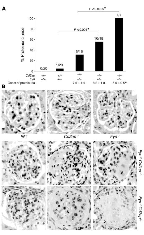
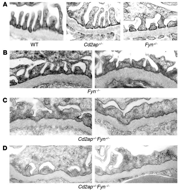
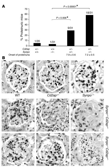
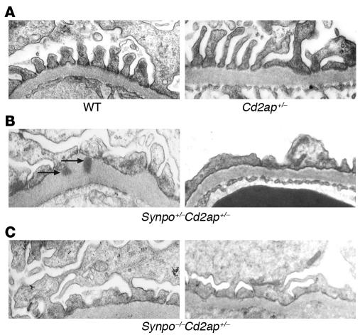
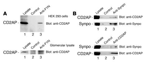
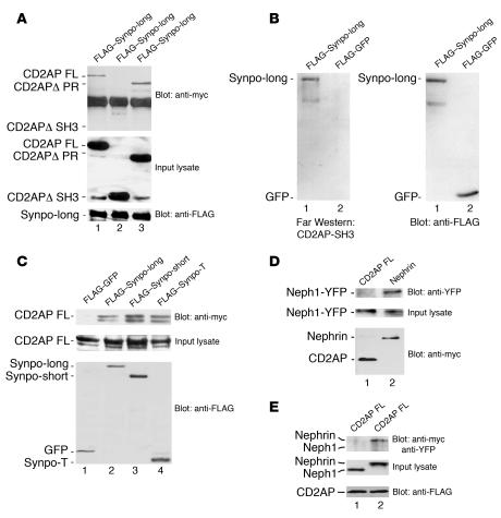
Focal segmental glomerulosclerosis (FSGS) is the most common primary glomerular diagnosis resulting in end-stage renal disease. Defects in several podocyte proteins have been implicated in the etiology of FSGS, including podocin, alpha-actinin-4, CD2-associated protein (CD2AP), and TRPC6. Despite our growing understanding of genes involved in the pathogenesis of focal segmental sclerosis, the vast majority of patients with this disease, even those with a familial linkage, lack a clear genetic diagnosis. Here, we tested whether combinations of genetic heterozygosity (bigenic heterozygosity) that alone do not result in clinical kidney disease could function together to enhance susceptibility to glomerular damage and FSGS. Combinations of Cd2ap heterozygosity and heterozygosity of either synaptopodin (Synpo) or Fyn proto-oncogene (Fyn) but not kin of IRRE like 1 (Neph1) resulted in spontaneous proteinuria and in FSGS-like glomerular damage. These genetic interactions were also reflected at a functional level, as we found that CD2AP associates with Fyn and Synpo but not with Neph1. This demonstrates that bigenic heterozygosity can lead to FSGS and suggests that combined mutations in 2 or multiple podocyte genes may be a common etiology for glomerular disease.


Figures

Figure 1
Figure 1. CD2AP haploinsufficiency enhances proteinuria in Fyn-deficient mice.
(A) Mice were tested monthly for proteinuria. The incidence of proteinuria is depicted as both percentages and absolute numbers for the indicated mouse genotypes followed to up to 12 months of age. The onset of proteinuria is shown below for each genotype in months ± SEM. *Statistical significance. (B) Sections of formalin-fixed kidney cortex from 5- to 8-month-old mice were stained with H&E and with PAS. In Fyn–/– mice, most glomeruli were normal (top right). Representative glomeruli from Fyn+/–Cd2ap+/– mice (middle row) or Fyn–/–Cd2ap+/– mice (bottom row) taken 2 months after the onset of proteinuria reveal mesangial hypercellularity and segmental sclerosis or global sclerosis in some glomeruli. Magnification, ×400.
Figure 2
Figure 2. Electron micrograph images from combined heterozygous mice between Cd2ap and Fyn.
(A) Representative electron microscopic images showing normal foot process architecture in wild-type (left panel), Cd2ap+/– (middle panel), and Fyn+/– (right panel) podocytes. (B) Electron micrographs from proteinuric Fyn–/– mice revealed only some coarsening of the foot processes. (C and D) Fyn+/–Cd2ap+/– mice (C) and Fyn–/–Cd2ap+/– mice (D) exhibited foot process effacement in some but not all areas. Magnification, ×12,000.
Figure 3
Figure 3. Bigenic Cd2ap and Synpo heterozygosity lead to proteinuria and FSGS.
(A) Mice were screened monthly for proteinuria. The incidence of proteinuria is depicted as both percentages and absolute numbers for the indicated mouse genotypes followed for up to 12 months. The onset of proteinuria is shown below for each proteinuric genotype in months ± SEM and did not differ among genotypes. *Statistical significance. (B) Sections of formalin-fixed kidney cortex from 6- to 9-month-old mice were stained with H&E and with PAS. In wild-type, Synpo–/–, and CD2AP+/– mice, nearly all glomeruli were normal (top panel). Representative glomeruli from Synpo+/–Cd2ap+/– mice (middle row) or Synpo–/–Cd2ap+/– mice (bottom row) 2 months after the onset of proteinuria showed mesangial hypercellularity and segmental or global sclerosis in some but not all glomeruli. Magnification, ×400.
Figure 4
Figure 4. Electron micrograph images from combined heterozygous mice between Cd2ap and Synpo.
(A) Representative electron microscopic images showing normal foot process architecture in wild-type (left panel) or Cd2ap+/– mice (right panel). (B) Electron micrographs from proteinuric Synpo+/–CD2AP+/– showed extensive foot process effacement as well as subepithelial deposits (arrows). (C) Electron micrographs from proteinuric Synpo–/–CD2AP+/– showed findings similar to those shown in B, with foot process effacement. Magnification, ×12,000.
Figure 5
Figure 5. CD2AP forms an endogenous complex with Fyn and Synpo.
(A) Endogenous CD2AP interacts with endogenous Fyn in cells (upper panel) and rat glomeruli (lower panel). Lysates were prepared from HEK 293 cells or isolated rat glomeruli, respectively (input lysate, 25 μl out of 800 ml; lane 1). Immunoprecipitates were prepared with a control antibody (rabbit serum, lane 2) or with polyclonal antisera to Fyn (lane 3). Immunoblotting was performed with a polyclonal antibody to CD2AP. (B) Endogenous CD2AP interacts with endogenous Synpo in podocytes. Lysates were prepared from differentiated podocytes. Immunoprecipitates were prepared with a control antibody and anti-Synpo (upper panel) or anti-CD2AP (lower panel) antibodies and immunoblotted with anti-Synpo or anti-CD2AP.
Figure 6
Figure 6. CD2AP SH3 domains interact directly with Synpo, but CD2AP does not interact with Neph1.
(A) FLAG-tagged Synpo (Synpo-long) was coexpressed with myc-tagged proteins, including full-length CD2AP (lane 1, CD2AP FL), CD2AP lacking the SH3 domains (lane 2, ΔSH3 domains), and CD2AP lacking the proline-rich domains (lane 3, ΔPR domains). Anti-FLAG immunoprecipitates were immunoblotted with an anti-myc antibody (upper panel). The expression of wild-type and mutated CD2AP proteins (input lysate; anti-myc) and Synpo is shown in the middle and lower panels, respectively. (B) FLAG-tagged Synpo (lane 1, FLAG–Synpo-long) and FLAG-tagged GFP (lane 2) were overexpressed, and anti-FLAG immunoprecipitates were blotted with recombinant, biotinylated GST-CD2AP fusion protein containing the 3 SH3 domains (left panel) or immunoblotted with anti-FLAG antibody (right panel). (C) FLAG-tagged GFP (lane 1, control), FLAG-tagged Synpo-long (lane 2), FLAG-tagged Synpo-short (lane 3), and FLAG-tagged Synpo-T (lane 4) were coexpressed with myc-tagged full-length CD2AP. Immunoprecipitates were prepared with an anti-FLAG antibody and immunoblotted with an anti-myc antibody (upper panel). (D) Neph1–yellow fluorescent protein (Neph1-YFP) was coexpressed with myc-tagged full-length CD2AP (lane 1) or myc-tagged nephrin (lane 2). Anti-myc immunoprecipitates were immunoblotted with anti-YFP antibody (upper panel). The expression of Neph1-YFP input lysate and myc-tagged nephrin or CD2AP controls is shown in the middle and lower panels, respectively. (E) Neph1-YFP (lane 1) and myc-nephrin (lane 2) were coexpressed with full-length FLAG-tagged CD2AP. Immunoprecipitates of FLAG-CD2AP were immunoblotted with anti-myc and anti-YFP antibodies. Neph1-YFP or myc-nephrin input lysates (anti-myc, anti-YFP) and CD2AP expression are demonstrated in the middle and lower panels, respectively.

References

    1. Kitiyakara C., Kopp J.B., Eggers P. Trends in the epidemiology of focal segmental glomerulosclerosis. Semin. Nephrol. 2003;23:172–182. - PubMed
    1. Pollak M.R. The genetic basis of FSGS and steroid-resistant nephrosis. Semin. Nephrol. 2003;23:141–146. - PubMed
    1. Fogo A.B. Animal models of FSGS: lessons for pathogenesis and treatment. Semin. Nephrol. 2003;23:161–171. - PubMed
    1. Boute N., et al. NPHS2, encoding the glomerular protein podocin, is mutated in autosomal recessive steroid-resistant nephrotic syndrome. Nat. Genet. 2000;24:349–354. - PubMed
    1. Kaplan J.M., et al. Mutations in ACTN4, encoding alpha-actinin-4, cause familial focal segmental glomerulosclerosis. Nat. Genet. 2000;24:251–256. - PubMed

Publication types