N-methyl-D-aspartate receptor subunit phenotypes of vagal afferent neurons in nodose ganglia of the rat
- PMID: 16628619
- PMCID: PMC2834225
- DOI: 10.1002/cne.20955
N-methyl-D-aspartate receptor subunit phenotypes of vagal afferent neurons in nodose ganglia of the rat
Abstract
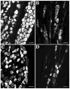
Most vagal afferent neurons in rat nodose ganglia express mRNA coding for the NR1 subunit of the heteromeric N-methyl-D-aspartate (NMDA) receptor ion channel. NMDA receptor subunit immunoreactivity has been detected on axon terminals of vagal afferents in the dorsal hindbrain, suggesting a role for presynaptic NMDA receptors in viscerosensory function. Although NMDA receptor subunits (NR1, NR2B, NR2C, and NR2D) have been linked to distinct neuronal populations in the brain, the NMDA receptor subunit phenotype of vagal afferent neurons has not been determined. Therefore, we examined NMDA receptor subunit (NR1, NR2B, NR2C, and NR2D) immunoreactivity in vagal afferent neurons. We found that, although the left nodose contained significantly more neurons (7,603), than the right (5,978), the proportions of NMDA subunits expressed in the left and right nodose ganglia were not significantly different. Immunoreactivity for NMDA NR1 subunit was present in 92.3% of all nodose neurons. NR2B immunoreactivity was present in 56.7% of neurons; NR2C-expressing nodose neurons made up 49.4% of the total population; NR2D subunit immunoreactivity was observed in just 13.5% of all nodose neurons. Double labeling revealed that 30.2% of nodose neurons expressed immunoreactivity to both NR2B and NR2C, whereas NR2B and NR2D immunoreactivities were colocalized in 11.5% of nodose neurons. NR2C immunoreactivity colocalized with NR2D in 13.1% of nodose neurons. Our results indicate that most vagal afferent neurons express NMDA receptor ion channels composed of NR1, NR2B, and NR2C subunits and that a minority phenotype that expresses NR2D also expresses NR1, NR2B, and NR2C.
Copyright 2006 Wiley-Liss, Inc.
Figures


References
-
- Aicher SA, Sharma S, Pickel VM. N-methyl-D-aspartate receptors are present in vagal afferents and their dendritic targets in the nucleus tractus solitarius. Neurosci. 1999;91:119–132. - PubMed
-
- Akazawa C, Shigemoto R, Bessho Y, Nakanishi S, Mizuno N. Differential expression of five N-methyl-D-aspartate receptor subunit mRNAs in the cerebellum of developing and adult rats. J Comp Neurol. 1994;347:150–160. - PubMed
-
- Allchin RE, Batten TF, McWilliam PN, Vaughan PF. Electrical stimulation of the vagus increases extracellular glutamate recovered from the nucleus tractus solitarii of the cat by in vivo microdialysis. Exp Physiol. 1994;79:265–268. - PubMed
-
- Andres ME, Gysling K, Araneda S, Venegas A, Bustos G. NMDA-NR1 receptor subunit mRNA expression in rat brain after 6-OH-dopamine induced lesions: a non-isotopic in situ hybridization study. J Neurosci Res. 1996;46:375–384. - PubMed
-
- Andresen MC, Mendelowitz D. Sensory afferent neurotransmission in caudal nucleus tractus solitarius--common denominators. Chem Senses. 1996;21:387–395. - PubMed
Publication types
MeSH terms
Substances
Grants and funding
LinkOut - more resources
Full Text Sources

