. 2006 May;60(3):630-43.
doi: 10.1111/j.1365-2958.2006.05135.x.

Flk prevents premature secretion of the anti-sigma factor FlgM into the periplasm

Affiliations

Flk prevents premature secretion of the anti-sigma factor FlgM into the periplasm

Phillip Aldridge et al. Mol Microbiol. 2006 May.

Erratum in

  • Mol Microbiol. 2006 Jul;61(1):269

Abstract

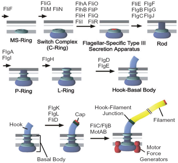
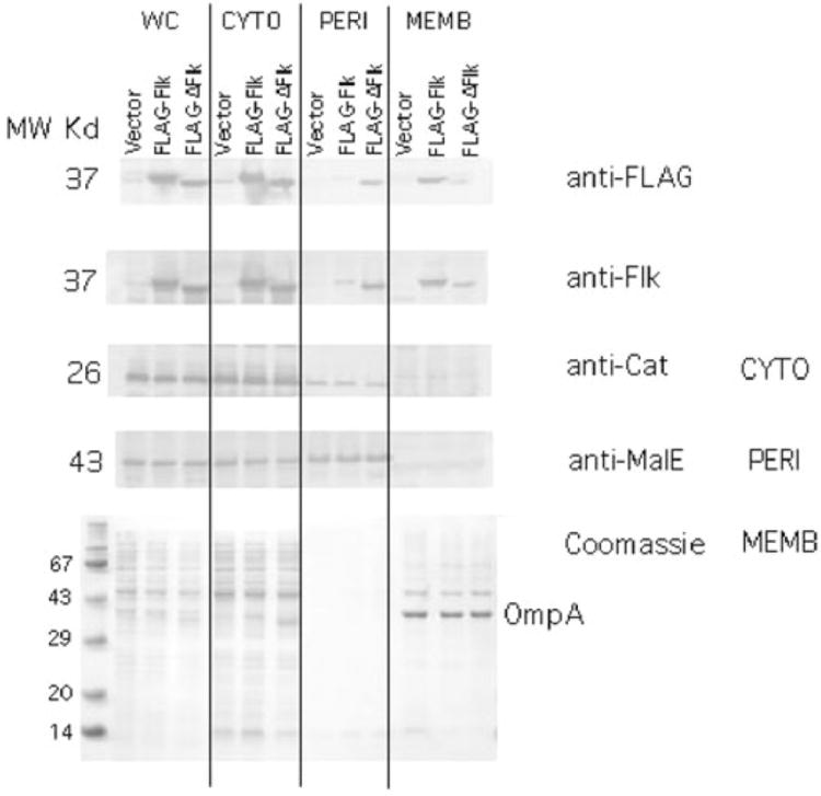
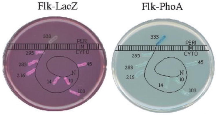
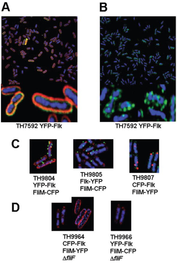
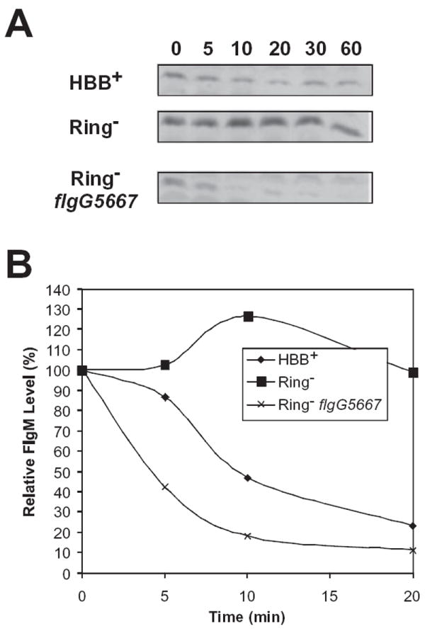
The flk locus of Salmonella typhimurium was identified as a regulator of flagellar gene expression in strains defective in P- and l-ring formation. Flk acts as a regulator of flagellar gene expression by modulating the protein levels of the anti-sigma28 factor FlgM. Evidence is presented which suggests that Flk is a cytoplasmic-facing protein anchored to the inner membrane by a single, C-terminal transmembrane-spanning domain (TMS). The specific amino acid sequence of the TMS is not essential for Flk activity, but membrane anchoring is essential. Membrane fractionation and visualization of protein fusions of green fluorescent protein derivatives to Flk suggested that the Flk protein is present in the membrane as punctate spots in number that are much greater than the number of flagellar basal structures. The turnover of the anti-sigma28 factor FlgM was increased in flk mutant strains. Using FlgM-beta-lactamase fusions we show the increased turnover of FlgM in flk null mutations is due to FlgM secretion into the periplasm where it is degraded. Our data suggest that Flk inhibits FlgM secretion by acting as a braking system for the flagellar-associated type III secretion system. A model is presented to explain a role for Flk in flagellar assembly and gene regulatory processes.


Figures

Fig. 1
Fig. 1
The flagellar assembly pathway of Salmonella typhimurium.
Fig. 2
Fig. 2
Cellular fractionation of the Flk protein. Plasmids pJK462 and pJK463 express Flk or FLAG–flk from the T7 promoter respectively. These plasmids and a vector control, pJK447 were placed in strain TH4657 that harbours T7 RNA polymerase under control of the lac promoter (Table 2). Following induction of T7 RNA polymerase, the whole cells (WC) were separated into cytoplasmic (CYTO), periplasmic (PERI) and membrane (MEMB) fractions (Experimental procedure). The proteins chloramphenicol acetyl transferase (Cat), maltose binding protein (MalE) and outer membrane protein A (OmpA) were used as controls for proteins known to be cytoplasmic, periplasmic and membrane-associated respectively. The fractions were run on SDS-PAGE and analysed by Western blot with anti-FLAG, anti-Flk, anti-Cat, anti-MalE antibodies. MW, molecular weight.
Fig. 3
Fig. 3
Flk is inserted into the membrane by a C-terminal hydrophobic tail. A diagram of the membrane topology of the Flk protein in the inner (cytoplasmic) membrane based on β-galactosidase (LacZ) and alkaline phosphatase (PhoA) fusion activities. The protein is represented as being primarily cytoplasmic (CYTO) anchored in the inner membrane with a C-terminal hydrophobic transmembrane segment that extends to the periplasm (PERI). Translational fusions of various proteins of the N-terminus of Flk were fused to LacZ (left) and PhoA (right) and assayed on indicator media. The numbers indicate the amino acid fusion junction of the LacZ or PhoA reporter to the specified amino acid position in Flk. On MacConkey-lactose medium (left) Lac+ cells are red and Lac cells are white. On medium containing the chromogenic substrate for PhoA, Xpho (right) PhoA+ cells are blue and PhoA cells are white. All LacZ fusions were Lac+ except the fusion at position 333. Conversely, all PhoA fusions were PhoA, except the fusion at position 333.
Fig. 4
Fig. 4
Flk appears as discrete spots in the cell membrane. A. Expression of YFP–Flk is associated with the membrane fraction as indicated by the membrane stain FM 4-64. B. Visualization of YFP–Flk in the absence of membrane stain indicates that it forms discrete spots on the cell membrane. C. Strains TH9804 expressing an N-terminal YFP fusion to Flk (YFP–Flk) and a CFP-labelled HBB (FliM–CFP), TH9805 expressing a C-terminal YFP fusion to Flk (Flk–YFP) and TH9807 expressing an N-terminal CFP fusion to Flk (CFP–Flk) and a YFP-labelled HBB (FliM–YFP). D. Strains TH9964 and TH9966 express CFP–Flk and FliM–YFP or YFP–Flk and FliM–CFP respectively, but are also deleted for the fliF gene, which is required for FliM assembly into HBB structures. Strain TH9964 was stained either with DAPI and the CFP–Flk visualized in red (TH9964, left photo) or with FM 4-64 and CFP–Flk visualized in blue (TH9964 right photo). TH9966 was stained with DAPI and YFP–FLK visualized in red. For TH9964 or TH9966, FliM–YFP or FliM–CFP, respectively, was not detected.
Fig. 5
Fig. 5
FlgM is unstable in the absence of Flk. A. Immunoblot analysis of FlgM protein at different time points following the addition of the translation inhibitor spectinomycin. Cellular FlgM turns over more rapidly in a strain with a functional hook-basal body (TH4803, HBB+) because of FlgM secretion. In a strain deleted for the P- and l-rings (TH7025, Ring), FlgM accumulates in the cell and is relatively stable. A null mutation in flk (TH7026) results in a sharp decrease in FlgM stability in the ring mutant strain. B. A graph representation of the data in (A).
Fig. 6
Fig. 6
FlgM–Bla is secreted into the periplasm in flk ΔflgHI strains. Immunoblot analysis of FlgM and a FlgM–Bla fusion in cellular and periplasmic fractions (see Experimental procedures). Strains were assayed for the ability to secrete either FlgM or a chromosomal FlgM–Bla fusion into the periplasm. Lanes 1 and 2, TH4803 (HBB+flgM+) and TH7025 (RingflgM+), respectively, has FlgM in the cytoplasm, but not in the periplasm. Lanes 3–5, TH9480 (HBB+flgM–bla), TH9481 (RingflgM–bla) and TH9486 (HBB+ Δflk flgM–bla), respectively, has FlgM–Bla in the cytoplasm, but not in the periplasm. Lane 6, TH9488 (Ring Δflk flgM–bla) and has FlgM–Bla secreted into the periplasm. Maltose binding protein (MBP) was used as a periplasmic protein control.

References

    1. Aizawa S-I. Flagellar assembly in Salmonella typhimurium. Mol Microbiol. 1996;20:1–4. - PubMed
    1. Aldridge P, Jenal U. Cell cycle-dependent degradation of a flagellar motor component requires a novel-type response regulator. Mol Microbiol. 1999;32:379–391. - PubMed
    1. Aldridge P, Karlinsey J, Hughes KT. The type III secretion chaperone FlgN regulates flagellar assembly via a negative feedback loop containing its chaperone substrates FlgK and FlgL. Mol Microbiol. 2003;49:1333–1345. - PubMed
    1. Benoit S, Abaibou H, Mandrand-Berthelot MA. Topological analysis of the aerobic membrane-bound formate dehydrogenase of Escherichia coli. J Bacteriol. 1998;180:6625–6634. - PMC - PubMed
    1. Boyd D, Schierle C, Beckwith J. How many membrane proteins are there? Protein Sci. 1998;7:201–205. - PMC - PubMed

Publication types

MeSH terms