. 2007 Jan 1;61(1):127-35.
doi: 10.1016/j.biopsych.2006.03.002. Epub 2006 Apr 21.

Acute stress and nicotine cues interact to unveil locomotor arousal and activity-dependent gene expression in the prefrontal cortex

Affiliations

Acute stress and nicotine cues interact to unveil locomotor arousal and activity-dependent gene expression in the prefrontal cortex

Craig A Schiltz et al. Biol Psychiatry. .

Abstract

Background: This study examines the interactive effects of acute stress and nicotine-associated contextual cues on locomotor activity and activity-dependent gene expression in subregions of the prefrontal cortex.

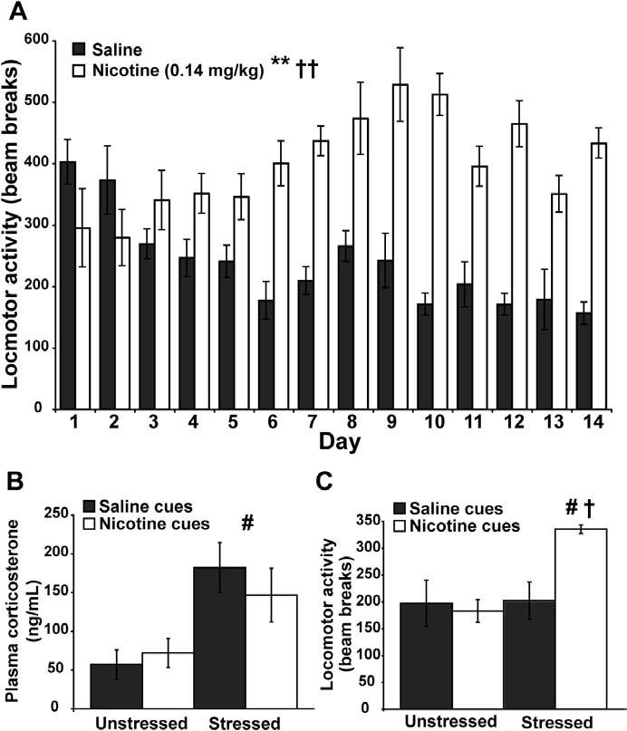
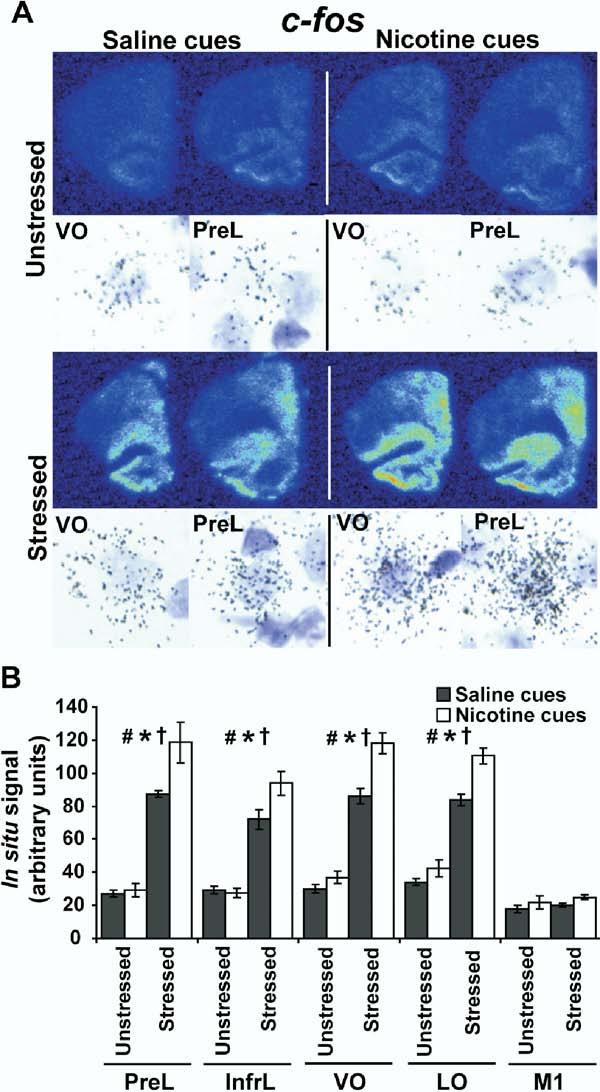
Methods: Locomotor activity of rats was measured in a context associated with either low-dose nicotine or saline administration with or without 5 minutes of pre-exposure to ferrets, a nonphysical stressor. After 45 minutes in the test environment, plasma corticosterone levels and mRNA levels of the immediate-early genes Arc, NGFI-B, and c-Fos in prefrontal and primary motor cortical subregions were measured.

Results: Stress alone increased plasma corticosterone and prefrontal cortex gene expression. Low-dose nicotine cues had no effect on corticosterone levels nor did they elicit conditioned motor activation, and they caused minor elevations in gene expression. Stress and low-dose nicotine cues, however, interacted to elicit conditioned motor activation and further increases in early response gene expression in prefrontal but not in the primary motor cortical subregions.

Conclusions: Stress interacts with nicotine-associated cues to uncover locomotor arousal, a state associated with prefrontal neuronal activation and immediate early gene expression. Thus, in nicotine-experienced individuals, stress may be an important determinant of subjective reactivity and prefrontal cortex activation that occurs in response to nicotine-associated cues.


Figures

Figure 1
Figure 1
Schematic diagrams of the experimental paradigm and the brain regions analyzed for gene expression. (A)The experimental paradigm consisted of a 14-day training period in which all rats received one of two daily injections in two distinct environments (see Methods and Materials). Loco-motor activity was measured in context A over the training phase of the study. On the test day, 3 days following the final day of training, rats were reintroduced to context A with or without 5 minutes of ferret exposure immediately prior to reentry into context A. Locomotor activity was measured in context A for 45 minutes, after which time rats were sacrificed and trunk blood and brains were taken for plasma corticosterone assay and in situ hybridization analysis of immediate early gene expression in the pre-frontal and the primary motor cortices. (S.C. = saline cues, N.C. = nicotine cues). (B)The brain regions analyzed for activity-dependent gene expression via in situ hybridization included the prelimbic cortex (PreL), infralimbic cortex (InfrL), ventral orbital prefrontal cortex (VO), lateral orbital prefrontal cortex (LO), and the primary motor cortex (M1). Numbers represent distance from bregma in millimeters. S.C., saline cues; N.C., nicotine cues; PreL, prelimbic cortex; InfrL, infralimbic cortex; VO, ventral orbital prefrontal cortex; LO, lateral orbital prefrontal cortex; M1, primary motor cortex. (Adapted with permission from Paxinos and Watson 1998).
Figure 2
Figure 2
Behavioral and corticosterone data. (A)Daily locomotor activity in context A over the 14-day training period. Nicotine significantly potentiated locomotor activity over the training period (n = 8 per group, **p < .001). There was also a significant treatment × day interaction (††p < .01). (B)Plasma corticosterone levels on the test day. While cues did not have an effect on plasma corticosterone levels, rats exposed to predator stress exhibited significantly higher levels of plasma corticosterone than unstressed rats (n = 4 per group, #p < .05). (C)Locomotor activity data on the test day. There was a main effect of stress, but not of cues, on locomotor activity on the test day (n = 4 per group, #p < .05). There was also a significant interaction between stress and cues on locomotor activity, which was evident by increased motor activity in the group of rats stressed and exposed to nicotine cues (n = 4 per group, †p < .05).
Figure 3
Figure 3
Expression of arc in the rat brain in response to stress and nicotine cues. (A)Representative psuedocolor phosphorescence autoradiograms of coronal, right hemisphere sections from each of the experimental groups hybridized with a probe against arc. In each panel, the upper left pair of sections is from the unstressed/saline cues group, the upper right pair are from the unstressed/nicotine cues group, the lower left pair are from the stressed/saline cues group, and the lower right pair are from the stressed/nicotine cues group. Directly below each autoradiogram is a brightfield photomicrograph illustrating silver grain accumulation over an individual, large, lightly Nissl stained nuclei from the ventral orbital cortex (VO) and prelimbic cortex (PreL) taken from autoradiographic emulsions for each of the groups. This pattern of silver grain accumulation is consistent with a neuronal expression pattern for arc and was consistent across the prefrontal subregions analyzed. (B)Graphical representation of densitometric analysis for the prelimbic (PreL), infralimbic (InfrL), ventral orbital (VO), lateral orbital (LO), and primary motor (M1) cortices in each of the experimental groups for arc expression (n = 4 per group). There was a main effect of stress on arc expression in the PreL [F(1,12) = 118.611, p < .0001], InfrL [F(1,12) = 104.596, p < .0001], VO [F(1,12) = 235.464, p < .0001], LO [F(1,12) = 320.311, p < .0001], and the M1 [F(1,12) = 8.791, p = .0118] (#p < .05). There was also a main effect of cues on arc expression in the PreL [F(1,12) = 7.349, p = .0189], InfrL [F(1,12) = 8.725, p = .0121],VO[F(1,12)=16.157,p=.0017],andLO[F(1,12)=28.662,p=.0002],but not in the M1 [F(1,12) = 3.657, p = .0800] (*p < .05). In addition, stress and cues interacted to potentiate arc expression in the PreL [F(1,12) = 6.125, p = .0292], InfrL [F(1,12) = 8.335, p = .0136], VO [F(1,12) = 5.898, p = .0318], and LO [F(1,12) = 10.556, p = .0070] but not in the M1 [F(1,12) = .033, p = .8592] (†p < .05). VO, ventral orbital prefrontal cortex; PreL, prelimbic cortex; InfrL, infralimbic cortex; LO, lateral orbital prefrontal cortex; M1, primary motor cortex.
Figure 4
Figure 4
Expression of ngfi-b in the rat brain in response to stress and nicotine cues. (A)Representative psuedocolor phosphorescence autoradio-grams of coronal, right hemisphere sections from each of the experimental groups hybridized with a probe against ngfi-b (same layout as Figure 3). Brightfield photomicrographs illustrate silver grain accumulation over individual, large, lightly Nissl stained nuclei from the VO and PreL taken from autoradiographic emulsions for each of the groups. This pattern of silver grain accumulation is consistent with a neuronal expression pattern for ngfi-b and was consistent across the prefrontal subregions analyzed. (B)Graphical representation of densitometric analysis of ngfi-b expression in brain regions (n = 4 per group). There was a main effect of stress on ngfi-b expression in the PreL [F(1,12) = 84.448, p < .0001], InfrL [F(1,12) = 54.142, p < .0001], VO [F(1,12) = 108.905, p < .0001], and the LO [F(1,12) = 55.016, p < .0001] but not in the M1 [F(1,12) = 4.136, p = .0647] (#p < .05). There was also a main effect of cues on ngfi-b expression in the PreL [F(1,12) = 14.481, p = .0025], the InfrL [F(1,12) = 11.976, p = .0047], the VO [F(1,12) = 8.9466, p = .0113], the LO [F(1,12) = 7.128, p = .0204], and the M1 [F(1,12) = 12.112, p = .0045] (*p < .05).VO,ventralorbitalprefrontalcortex;PreL,prelimbiccortex;InfrL,infralimbic cortex; LO, lateral orbital prefrontal cortex; M1, primary motor cortex.
Figure 5
Figure 5
Expression of c-fos in the rat brain in response to stress and nicotine cues. (A)Representative psuedocolor phosphorescence autoradiograms of coronal, right hemisphere sections from each of the experimental groups hybridized with a probe against c-fos (same layout as Figure 3). Brightfield photomicrographs illustrate silver grain accumulation over individual, large, lightly Nissl stained nuclei from the VO and PreL taken from autoradiographic emulsions for each of the groups. This pattern of silver grain accumulation is consistent with a neuronal expression pattern for c-fos and was consistent across the prefrontal subregions analyzed. (B)Graphical representation of densitometric analysis for the PreL, InfrL, VO, LO, and M1 cortices in each of the experimental groups for c-fos expression (n = 4 per group). There was a main effect of stress on c-fos expression in PreL [F(1,12) = 127.873, p < .0001], InfrL [F(1,12) = 117.443, p < .0001], VO [F(1,12) = 241.081, p < .0001], and LO [F(1,12) = 209.024, p < .0001] but not M1 cortices [F(1,12) = 1.219, p = .2912] (#p < .05). There was also a main effect of cues on c-fos expression in the PreL [F(1,12) = 6.286, p = .0276], InfrL [F(1,12) = 4.037, p = .0675], VO [F(1,12) = 19.326, p = .0009], and LO cortices [F(1,12) = 18.377, p = .0011] but not the M1 cortex [F(1,12) = 3.150, p= .1013]) (*p < .05). In addition, stress and cues interacted to potentiate c-fos expression in the PreL [F(1,12) = 4.859, p = .0478], InfrL [F(1,12) = 5.362, p = .0391], VO [F(1,12) = 8.251, p = .0140], and LO cortices [F(1,12) = 5.082, p= .0437] but not in the M1 cortex [F(1,12) = .029, p = .8669] (†p< .05). VO, ventral orbital prefrontal cortex; PreL, prelimbic cortex; InfrL, infralimbic cortex; LO, lateral orbital prefrontal cortex; M1, primary motor cortex.

Similar articles

Cited by

References

    1. Adamec R. Does long term potentiation in periacqueductal gray (PAG) mediate lasting changes in rodent anxiety-like behavior (ALB) produced by predator stress?-Effects of low frequency stimulation (LFS) of PAG on place preference and changes in ALB produced by predator stress. Behav Brain Res. 2001;120:111–135. - PubMed
    1. Bassareo V, De Luca MA, Di Chiara G. Differential expression of motivational stimulus properties by dopamine in nucleus accumbens shell versus core and prefrontal cortex. J Neurosci. 2002;22:4709–4719. - PMC - PubMed
    1. Bassareo V, Di Chiara G. Differential influence of associative and nonassociative learning mechanisms on the responsiveness of prefrontal and accumbal dopamine transmission to food stimuli in rats fed ad libitum. J Neurosci. 1997;17:851–861. - PMC - PubMed
    1. Bevins RA, Besheer J, Pickett KS. Nicotine-conditioned locomotor activity in rats: Dopaminergic and GABAergic influences on conditioned expression. Pharmacol Biochem Behav. 2001;68:135–145. - PubMed
    1. Bevins RA, Palmatier MI. Nicotine-conditioned locomotor sensitization in rats: Assessment of the US-preexposure effect. Behav Brain Res. 2003;143:65–74. - PubMed

Publication types

MeSH terms