. 2006 Jun 1;15(11):1847-57.
doi: 10.1093/hmg/ddl107. Epub 2006 Apr 21.

In-frame deletion in a novel centrosomal/ciliary protein CEP290/NPHP6 perturbs its interaction with RPGR and results in early-onset retinal degeneration in the rd16 mouse

Affiliations

In-frame deletion in a novel centrosomal/ciliary protein CEP290/NPHP6 perturbs its interaction with RPGR and results in early-onset retinal degeneration in the rd16 mouse

Bo Chang et al. Hum Mol Genet. .

Abstract

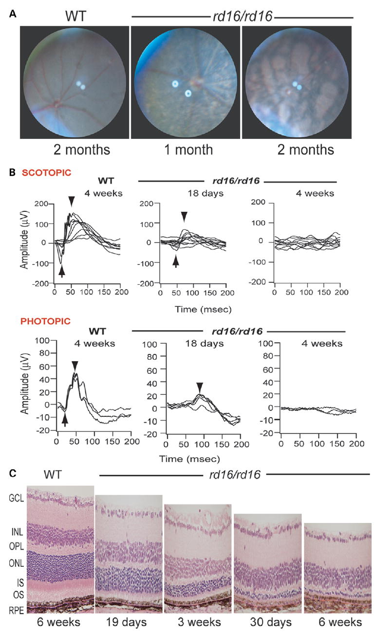
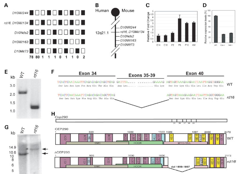
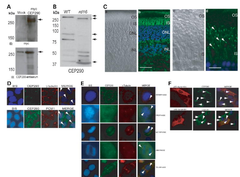
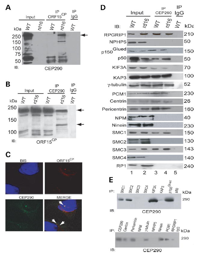
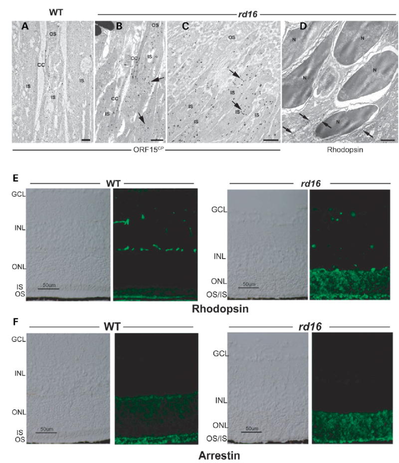
Centrosome- and cilia-associated proteins play crucial roles in establishing polarity and regulating intracellular transport in post-mitotic cells. Using genetic mapping and positional candidate strategy, we have identified an in-frame deletion in a novel centrosomal protein CEP290 (also called NPHP6), leading to early-onset retinal degeneration in a newly identified mouse mutant, rd16. We demonstrate that CEP290 localizes primarily to centrosomes of dividing cells and to the connecting cilium of retinal photoreceptors. We show that, in the retina, CEP290 associates with several microtubule-based transport proteins including RPGR, which is mutated in approximately 15% of patients with retinitis pigmentosa. A truncated CEP290 protein (DeltaCEP290) is detected in the rd16 retina, but in considerably reduced amounts; however, the mutant protein exhibits stronger association with specific RPGR isoform(s). Immunogold labeling studies demonstrate the redistribution of RPGR and of phototransduction proteins in the photoreceptors of rd16 retina. Our findings suggest a critical function for CEP290 in ciliary transport and provide insights into the mechanism of early-onset photoreceptor degeneration.


Conflict of interest statement

Conflict of Interest statement. The authors declare no conflict of interest.

Figures

Figure 1
Figure 1
Examination of the rd16 mouse retina. (A) Fundus photographs of WT C57BL/6J mouse and the rd16 homozygote mutants (rd16/rd16) demonstrating retinal degeneration at 1 month of age and at 2 months. (B) ERG responses of WT and mutant (rd16/rd16) mice under dark- (SCOTOPIC) and light- (PHOTOPIC) adapted conditions. Arrows indicate the A-wave and arrowheads the B-wave. (C) Histology of retina ofWT and rd16 homozygotesmice at indicated ages. RPE, retinal pigment epithelium; OS, outer segments; IS, inner segments; ONL, outer nuclear layer; OPL, outer plexiformlayer; INL, inner nuclear layer; GCL, ganglion cell layer.
Figure 2
Figure 2
Cep290 mutation in rd16. (A) Linkage cross-data: 165 back-cross progeny from the (rd16 × CAST/EiJ)F1 × rd16/rd16 were phenotyped for ERG phenotype and genotyped for the indicated microsatellite markers. Black boxes represent homozygosity for rd16-derived alleles and white boxes represent heterozygosity for rd16- and CAST-derived alleles. The number of animals sharing the corresponding haplotype is indicated below each column of squares. The order of marker loci was determined by minimizing the number of crossovers. The rd16 locus was inferred from the ERG phenotype of mice showing recombinations. (B) Genetic map of mouse chromosome 10 showing the rd16 critical region, which is syntenic to human chromosome 12q21.1. (C) Real-time RT-PCR analysis of BC004690 (Cep290, exons 27–48) in the retina of WT mice. The expression levels at different developmental stages were calculated as relative fold change with respect to embryonic day, E14, after normalization to Hprt levels. P, postnatal day. Each bar represents the mean ± SE (n = 6). (D) Real-time RT-PCR analysis of BC004690 in the retina of Crx−/ and Nrl−/− versus WT mice. The expression levels in the Crx−/− and Nrl−/− retina were calculated as percentage of the level in the WT mouse retina after normalization to Hprt levels. Each bar represents the mean ± SE (n = 6). (E) RT-PCR analysis (with F2–R2 primer set) of BC004690 using rd16 and WT retinal RNA. A 1.2 kb band is detected in rd16 compared with a 2.1 kb product in WT. DNA size markers are shown on the left (in kb). (F) BC004690 sequence in rd16 showing an in-frame deletion of 897 bp encompassing exons 35–39. Three-letter codes for amino acids were used. (G) Southern analysis of WT and rd16 DNA using an exon 34 probe. DNA was digested with EcoRV, which cuts the WT DNA five times between exons 34 and 40, whereas in the rd16 DNA, only three EcoRV sites remain. WT DNA gave the expected band of 10.6 kb, whereas with the rd16 DNA, a heavier band at ~15 kb (indicated by arrows) is seen. Molecular weight markers are in kilobases. (H) Schematic representation of the Cep290 gene and the CEP290 and ΔCEP290 proteins showing putative domains and motifs. CC, coiled-coil; KID, RepA/Re+ protein KID; P-loop, ATP-GTP-binding site motif A; spindle association (SA) domain; MYO-Tail, myosin tail homology domain.
Figure 3
Figure 3
Expression and localization of CEP290. (A) COS-1 cells were transfected with empty vector (mock) or a vector encoding full-length human CEP290 protein fused to a myc-tag. Cells were lysed and analyzed by immunoblotting (IB), using anti-myc (upper panel) or anti-CEP290 antiserum (lower panel). Arrows indicate specific bands. The immunoreactive band in the mock transfected lane (lower panel) is endogenous CEP290 protein. Pre-immune serum shows no signal (data not shown). (B) Immunoblots of protein extracts from WT (20 μg) and rd16 (200μg) retina were analyzed using CEP290 antibody. Arrows indicate the full length and predicted alternatively spliced products of CEP290. (C) Immunohistochemical analysis of WT mouse retina. The sections were incubated with the CEP290 antibody followed by secondary antibody incubation. (a) and (c) Nomarski image of the retinal sections. (b) and (d) Staining with the CEP290 antibody (green) reveals intense labeling of the connecting cilium (indicated by arrows). Labeling in the IS is also observed. Scale bar: 40 μm for (a), (b); 10 μm for (c), (d). (D) CEP290 (green) co-localizes with γ-tubulin (red; upper panel) and PCM1 (red; lower panel) at the centrosomes (arrows; merge) in IMCD-3 cells. Bisbenzimide (BIS) was used to stain the DNA. (E) CEP290 is associated with centrosomes during cell cycle. Synchronized HeLa cells were co-stained with antibodies against γ-tubulin (red) and CEP290 (green) and analyzed by confocal microscopy. Arrows indicate the centrosomal staining of CEP290 (merge) at all indicated stages of cell division. (F) IMCD-3 cells were transfected with p50-dynamitin expression construct. Cells were stained with p50 (red), CEP290 or γ-tubulin (green) antibodies. Arrows denote centrosomal CEP290 and γ-tubulin in untransfected cells, whereas arrowheads denote the localization of CEP290 and γ-tubulin to centrosomes in p50-overexpressing cells. Merge image shows blue nuclear staining.
Figure 4
Figure 4
CEP290 and Δ CEP290 associate with RPGR-ORF15 and other centrosomal/microtubule-associated proteins in the retina. (A, B) IP was performed using ORF15CP (A), CEP290 (B) antibodies or normal IgG from WT and rd16 retinal extract (200 μg each). The immunoprecipitated proteins were analyzed by IB using CEP290 (A) or ORF15CP (B) antibodies. Input lane contains 20% of the protein extract used for IP. Longer exposure of the blot in (A) shows an immunoreactive band for ΔCEP290 in rd16 input lane (data not shown). Molecular weight markers are shown in kilo Dalton. Asterisk indicates the faint full-length CEP290-immunoreactive band (290 kDa) immunoprecipitated from the WT retina using the ORF15CP antibody. Arrow in (A) points to the ΔCEP290 protein immunoprecipitated from rd16 retina using ORF15CP. Arrows in (B) indicate multiple RPGR-ORF15 isoforms recognized by the ORF15CP antibody (29). Less high molecular weight (120–220 kDa) RPGR-ORF15 isoforms are immunoprecipitated by the CEP290 antibody in rd16. (C) Immunocytochemistry using the CEP290 (green) and ORF15CP (red) antibodies shows co-localization of endogenous CEP290 and RPGR-ORF15 in IMCD-3 cells. Arrows indicate co-localization (Merge). (D) WT and rd16 retinal extracts were subjected to IP using the CEP290 antibody and analyzed by IB using indicated antibodies. Input lane represents 5% of the total protein extract used for IP. Molecular weight markers are shown in kilo Dalton. Lanes 1 and 2: input from WT and rd16 retinal extracts; 3 and 4: IP using the CEP290 antibody from WT and rd16, respectively; 5: IP with normal IgG from WT retina. (E) Reverse IP was performed by incubating protein extracts of WT retina with indicated antibodies for IP followed by IB using the CEP290 antibody. Molecular weight markers are shown in kilo Dalton.
Figure 5
Figure 5
Localization of RPGR-ORF15, rhodopsin and arrestin in rd16 retinas. (A–D) Immunogold EM of WT or rd16 retinas with indicated antibodies. Labeling with ORF15CP antibody showed a predominant connecting cilium (CC) staining of RPGR-ORF15 (A) as opposed to abnormal extensive labeling throughout the photoreceptor IS in the rd16 retina (B, C). Arrows indicate clusters of immunogold particles. Labeling of rhodopsin in the rd16 retina (D) is evident around the photoreceptor cell bodies (indicated by arrows) with no exclusive OS localization; N, nucleus. (E, F) Immunohistochemical analysis of the WT and rd16 retinas at P12, dissected under normal light/dark cycle, with antibodies against rhodopsin (E) or arrestin (F). As shown, both rhodopsin and arrestin are localized primarily in the OS of WT retina, whereas in rd16, rhodopsin and arrestin are also detected in the ONL and ISs of photoreceptors. OS in the rd16 retina degenerate at P12 and therefore are represented in conjunction with the inner segments (OS/IS). Scale bar: 50 μm.

Similar articles

Cited by

References

    1. Nigg EA. Centrosomes in Development and Disease. Wiley-VCH Verlag GmbH & Co; KGaA: 2004.
    1. Doxsey S. Re-evaluating centrosome function. Nat Rev Mol Cell Biol. 2001;2:688–698. - PubMed
    1. Beisson J, Wright M. Basal body/centriole assembly and continuity. Curr Opin Cell Biol. 2003;15:96–104. - PubMed
    1. Badano JL, Teslovich TM, Katsanis N. The centrosome in human genetic disease. Nat Rev Genet. 2005;6:194–205. - PubMed
    1. Young RW. Passage of newly formed protein through the connecting cilium of retina rods in the frog. J Ultrastruct Res. 1968;23:462–473. - PubMed

Publication types

MeSH terms

Associated data