

Characterization of the structure of RAMP1 by mutagenesis and molecular modeling
- PMID: 16632510
- PMCID: PMC1483116
- DOI: 10.1529/biophysj.106.084582
Characterization of the structure of RAMP1 by mutagenesis and molecular modeling
Abstract
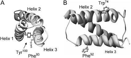
Receptor activity modifying proteins (RAMPs) are a family of single-pass transmembrane proteins that dimerize with G-protein-coupled receptors. They may alter the ligand recognition properties of the receptors (particularly for the calcitonin receptor-like receptor, CLR). Very little structural information is available about RAMPs. Here, an ab initio model has been generated for the extracellular domain of RAMP1. The disulfide bond arrangement (Cys27-Cys82, Cys40-Cys72, and Cys57-Cys104) was determined by site-directed mutagenesis. The secondary structure (alpha-helices from residues 29-51, 60-80, and 87-100) was established from a consensus of predictive routines. Using these constraints, an assemblage of 25,000 structures was constructed and these were ranked using an all-atom statistical potential. The best 1000 conformations were energy minimized. The lowest scoring model was refined by molecular dynamics simulation. To validate our strategy, the same methods were applied to three proteins of known structure; PDB:1HP8, PDB:1V54 chain H (residues 21-85), and PDB:1T0P. When compared to the crystal structures, the models had root mean-square deviations of 3.8 A, 4.1 A, and 4.0 A, respectively. The model of RAMP1 suggested that Phe93, Tyr100, and Phe101 form a binding interface for CLR, whereas Trp74 and Phe92 may interact with ligands that bind to the CLR/RAMP1 heterodimer.
Figures


Similar articles
-
Structure-function analysis of RAMP1-RAMP3 chimeras.Biochemistry. 2010 Jan 26;49(3):522-31. doi: 10.1021/bi9019093. Biochemistry. 2010. PMID: 20017504
-
Identification of N-terminal receptor activity-modifying protein residues important for calcitonin gene-related peptide, adrenomedullin, and amylin receptor function.Mol Pharmacol. 2008 Oct;74(4):1059-71. doi: 10.1124/mol.108.047142. Epub 2008 Jul 1. Mol Pharmacol. 2008. PMID: 18593822
-
Structure-function analysis of RAMP1 by alanine mutagenesis.Biochemistry. 2009 Jan 13;48(1):198-205. doi: 10.1021/bi801869n. Biochemistry. 2009. PMID: 19072332
-
Structure-function relationships of the N-terminus of receptor activity-modifying proteins.Br J Pharmacol. 2010 Mar;159(5):1059-68. doi: 10.1111/j.1476-5381.2009.00541.x. Epub 2009 Dec 10. Br J Pharmacol. 2010. PMID: 20015292 Free PMC article. Review.
-
Heterodimers and family-B GPCRs: RAMPs, CGRP and adrenomedullin.Biochem Soc Trans. 2004 Nov;32(Pt 5):843-6. doi: 10.1042/BST0320843. Biochem Soc Trans. 2004. PMID: 15494030 Review.
Cited by
-
Targeting a family B GPCR/RAMP receptor complex: CGRP receptor antagonists and migraine.Br J Pharmacol. 2012 May;166(1):66-78. doi: 10.1111/j.1476-5381.2011.01633.x. Br J Pharmacol. 2012. PMID: 21871019 Free PMC article. Review.
-
Calcitonin gene-related peptide: physiology and pathophysiology.Physiol Rev. 2014 Oct;94(4):1099-142. doi: 10.1152/physrev.00034.2013. Physiol Rev. 2014. PMID: 25287861 Free PMC article. Review.
-
Crystal structure of the human receptor activity-modifying protein 1 extracellular domain.Protein Sci. 2008 Nov;17(11):1907-14. doi: 10.1110/ps.036012.108. Epub 2008 Aug 25. Protein Sci. 2008. PMID: 18725456 Free PMC article.
-
CGRP induction in cystic fibrosis airways alters the submucosal gland progenitor cell niche in mice.J Clin Invest. 2011 Aug;121(8):3144-58. doi: 10.1172/JCI41857. Epub 2011 Jul 18. J Clin Invest. 2011. PMID: 21765217 Free PMC article.
-
Dissection of functional residues in receptor activity-modifying proteins through phylogenetic and statistical analyses.Evol Bioinform Online. 2008 Apr 28;4:153-69. doi: 10.4137/ebo.s705. Evol Bioinform Online. 2008. PMID: 19204815 Free PMC article.
References
-
- Prinster, S. C., C. Hague, and R. A. Hall. 2005. Heterodimerization of g protein-coupled receptors: specificity and functional significance. Pharmacol. Rev. 57:289–298. - PubMed
-
- McLatchie, L. M., N. J. Fraser, M. J. Main, A. Wise, J. Brown, N. Thompson, R. Solari, M. G. Lee, and S. M. Foord. 1998. RAMPs regulate the transport and ligand specificity of the calcitonin-receptor-like receptor. Nature. 393:333–339. - PubMed
-
- Christopoulos, G., K. J. Perry, M. Morfis, N. Tilakaratne, Y. Gao, N. J. Fraser, M. J. Main, S. M. Foord, and P. M. Sexton. 1999. Multiple amylin receptors arise from receptor activity-modifying protein interaction with the calcitonin receptor gene product. Mol. Pharmacol. 56:235–242. - PubMed
-
- Christopoulos, A., G. Christopoulos, M. Morfis, M. Udawela, M. Laburthe, A. Couvineau, K. Kuwasako, N. Tilakaratne, and P. M. Sexton. 2003. Novel receptor partners and function of receptor activity-modifying proteins. J. Biol. Chem. 278:3293–3297. - PubMed
Publication types
MeSH terms
Substances
Grants and funding
LinkOut - more resources
Full Text Sources
Other Literature Sources
