. 2006 May 2;103(18):6829-34.
doi: 10.1073/pnas.0510711103. Epub 2006 Apr 21.

Binding of 5'-GTP to the C-terminal FeS cluster of the radical S-adenosylmethionine enzyme MoaA provides insights into its mechanism

Affiliations

Binding of 5'-GTP to the C-terminal FeS cluster of the radical S-adenosylmethionine enzyme MoaA provides insights into its mechanism

Petra Hänzelmann et al. Proc Natl Acad Sci U S A. .

Abstract

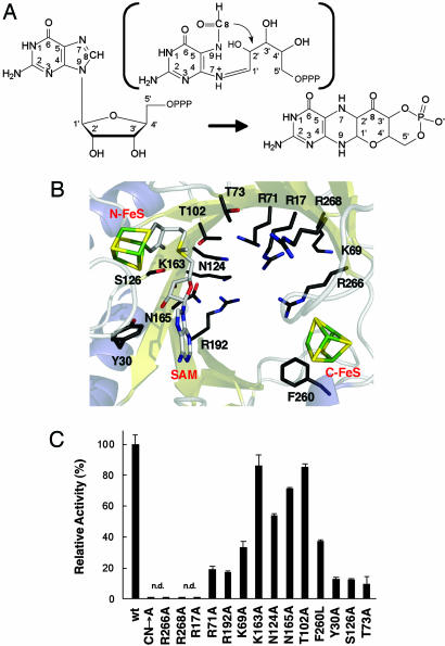
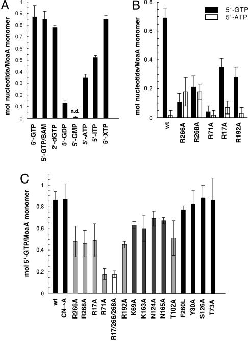
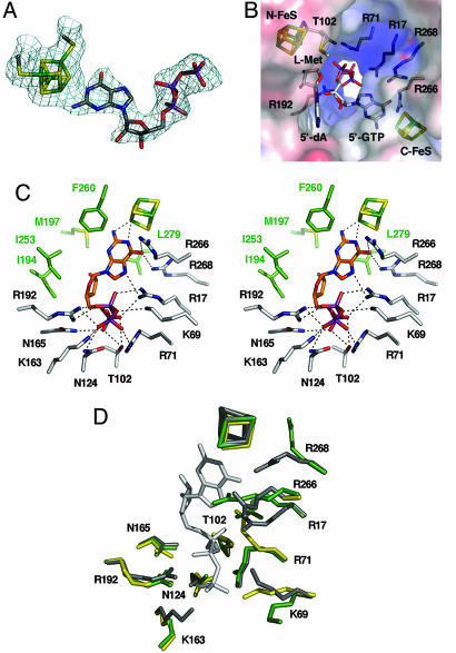
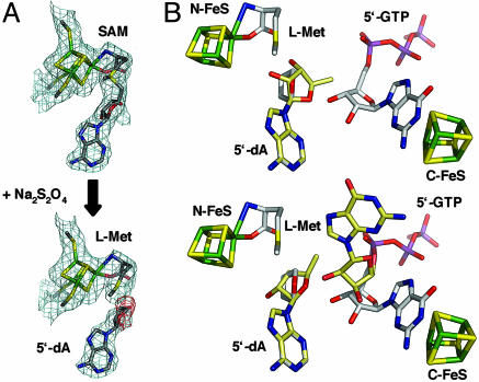
The first step in molybdenum cofactor biosynthesis, the conversion of 5'-GTP to precursor Z, an oxygen-sensitive tetrahydropyranopterin is catalyzed by the S-adenosylmethionine (SAM)-dependent enzyme MoaA and the accessory protein MoaC. This reaction involves the radical-initiated intramolecular rearrangement of the guanine C8 atom. MoaA harbors an N-terminal [4Fe-4S] cluster, which is involved in the reductive cleavage of SAM and generates a 5'-deoxyadenosyl radical (5'-dA*), and a C-terminal [4Fe-4S] cluster presumably involved in substrate binding and/or activation. Biochemical studies identified residues involved in 5'-GTP binding and the determinants of nucleotide specificity. The crystal structure of MoaA in complex with 5'-GTP confirms the biochemical data and provides valuable insights into the subsequent radical reaction. MoaA binds 5'-GTP with high affinity and interacts through its C-terminal [4Fe-4S] cluster with the guanine N1 and N2 atoms, in a yet uncharacterized binding mode. The tightly anchored triphosphate moiety prevents the escape of radical intermediates. This structure also visualizes the L-Met and 5'-dA cleavage products of SAM. Rotation of the 5'-dA ribose and/or conformational changes of the guanosine are proposed to bring the 5'-deoxyadenosyl radical into close proximity of either the ribose C2' and C3' or the guanine C8 carbon atoms leading to hydrogen abstraction.


Conflict of interest statement

Conflict of interest statement: No conflicts declared.

Figures

Fig. 1.
Fig. 1.
Synthesis of precursor Z and catalytic activities of MoaA active site variants. (A) Conversion of 5′-GTP to precursor Z. A proposed formamidopyrimidine-type intermediate is shown in parentheses. (B) Representation of the MoaA active site (PDB ID code 1TV8). Both [4Fe–4S] clusters, SAM, and conserved residues potentially involved in catalysis are displayed. Figs. 1B, 3, and 4 were created with pymol (www.pymol.org). (C) Synthesis of precursor Z determined by measurement of its stable oxidized derivative, compound Z, by reverse-phase HPLC. The activity of the WT protein (wt) was set to 100%. Error bars denote the SD from the mean of at least four independent experiments. n.d., not detected; CN→A, triple Cys→Ala variant of the N-terminal FeS cluster.
Fig. 2.
Fig. 2.
Determination of MoaA substrate-binding by equilibrium dialysis. (A) Nucleotide specificity. MoaA (75 μM) was equilibrated with the listed nucleotides (150 μM); n.d., not detected. (B) 5′-GTP/5′-ATP competition. WT MoaA (wt) and variants (75 μM) were dialyzed against equimolar concentrations (150 μM) of 5′-GTP (black bar) and 5′-ATP (white bar). (C) 5′-GTP binding. WT MoaA and variants (75 μM) were dialyzed against 5′-GTP (150 μM). Error bars denote the SD from the mean of at least four independent experiments. CN→A, triple Cys→Ala variant of the N-terminal FeS cluster; black bars, WT MoaA and residues not involved in 5′-GTP binding; light gray bars, residues important for 5′-GTP binding; dark gray bars, residues less important in 5′-GTP binding; white bar, R17/266/268A triple variant.
Fig. 3.
Fig. 3.
Structure of MoaA in complex with 5′-GTP. (A) 2FoFc map of the C-terminal [4Fe–4S] cluster with bound 5′-GTP contoured at one times the rms deviation. (B) Electrostatic potential (electropositive in blue, electronegative in red contoured at ±10 kT) surrounding the hydrophilic channel. [4Fe–4S] clusters, l-Met, 5′-dA, 5′-GTP, and residues important for 5′-GTP binding are displayed. (C) Stereoview of 5′-GTP interactions with surrounding active site residues (dashed lines). Carbon atoms of residues in hydrogen-bonding distance are in white, and carbon atoms of residues within a distance of 4 Å are in green. (D) Superposition of MoaA in complex with 5′-GTP (gray), without 5′-GTP (PDB ID code 1TV8; green) and the R17/266/268A variant (yellow).
Fig. 4.
Fig. 4.
Reductive cleavage of SAM and radical transfer from 5′-dA to 5′-GTP. (A) 2FoFc maps (blue; one rms deviation) of the N-terminal [4Fe–4S] cluster before (Upper) and after (Lower) reductive cleavage of SAM. FoFc map (red; −2.5 rms deviations) obtained with SAM as the model. (B) Radical transfer models. Carbon atoms of 5′-dA and 5′-GTP as present in the 5′-GTP/5′-dA structure are in white, and carbon atoms of postulated models are shown as pale yellow. (Upper) 5′-dA-ribose rotation model. (Lower) 5′-dA-ribose/guanosine rotation model.

Similar articles

Cited by

References

    1. Johnson D. C., Dean D. R., Smith A. D., Johnson M. K. Annu. Rev. Biochem. 2005;74:247–281. - PubMed
    1. Johnson M. K. Curr. Opin. Chem. Biol. 1998;2:173–181. - PubMed
    1. Beinert H., Holm R. H., Munck E. Science. 1997;277:653–659. - PubMed
    1. Beinert H. J. Biol. Inorg. Chem. 2000;5:2–15. - PubMed
    1. Flint D. H., Allen R. M. Chem. Rev. 1996;96:2315–2334. - PubMed

Publication types

MeSH terms

LinkOut - more resources