. 2006 May 2;103(18):7071-6.
doi: 10.1073/pnas.0509392103. Epub 2006 Apr 21.

Crystal structure and catalytic mechanism of the LPS 3-O-deacylase PagL from Pseudomonas aeruginosa

Affiliations

Crystal structure and catalytic mechanism of the LPS 3-O-deacylase PagL from Pseudomonas aeruginosa

Lucy Rutten et al. Proc Natl Acad Sci U S A. .

Abstract

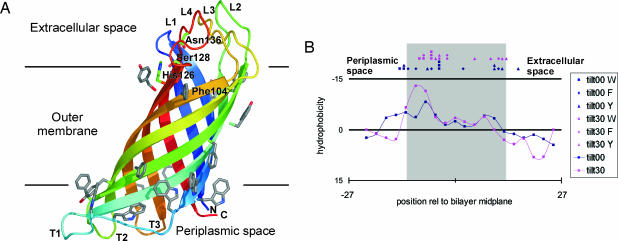
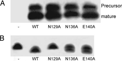
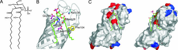
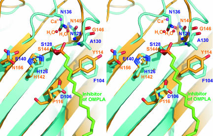
Pathogenic gram-negative bacteria can modify the lipid A portion of their lipopolysaccharide in response to environmental stimuli. 3-O-deacylation of lipid A by the outer membrane enzyme PagL modulates signaling through Toll-like receptor 4, leading to a reduced host immune response. We found that PagL is widely disseminated among gram-negative bacteria. Only four residues are conserved: a Ser, His, Phe, and Asn residue. Here, we describe the crystal structure of PagL from Pseudomonas aeruginosa to 2.0-A resolution. It consists of an eight-stranded beta-barrel with the axis tilted by approximately 30 degrees with respect to the lipid bilayer. The structure reveals that PagL contains an active site with a Ser-His-Glu catalytic triad and an oxyanion hole that comprises the conserved Asn. The importance of active site residues was confirmed in mutagenesis studies. Although PagL is most likely active as a monomer, its active site architecture shows high resemblance to that of the dimeric 12-stranded outer membrane phospholipase A. Modeling of the substrate lipid X onto the active site reveals that the 3-O-acyl chain is accommodated in a hydrophobic groove perpendicular to the membrane plane. In addition, an aspartate makes a hydrogen bond with the hydroxyl group of the 3-O-acyl chain, probably providing specificity of PagL toward lipid A.


Conflict of interest statement

Conflict of interest statement: No conflicts declared.

Figures

Fig. 1.
Fig. 1.
Folding and in vitro activity of recombinant PagL. (A) Coomassie-stained SDS/PAGE gel showing the heat modifiability of purified, refolded PagL. Samples were treated in sample buffer containing either 2% or 0.1% SDS and at room temperature (RT) or 100°C before electrophoresis. The positions of molecular mass standard proteins are shown at the right. (B) Purified N. meningitidis LPS was incubated in a detergent-containing buffer with or without refolded PagL and analyzed by Tricine-SDS/PAGE and staining with silver.
Fig. 2.
Fig. 2.
PagL structure and membrane orientation. (A) Ribbon representation of PagL. The N and C termini are labeled and colored blue and red, respectively, with gradient colors in between. The four extracellular loops are labeled L1–L4, and the three periplasmic turns are labeled T1–T3. Aromatic residues located at the presumed membrane boundaries are shown in gray, with nitrogen and oxygen atoms shown in blue and red, respectively. The only four completely conserved residues among PagL homologs are labeled. (B) Hydrophobicity profiles for the outward-facing PagL residues as a function of membrane position (periplasmic side left, extracellular side right) are shown as solid lines. Negative Σ(ΔG) values indicate regions that are more hydrophobic. The blue line and symbols present results for the positions with the β-barrel axis aligned along the membrane normal, whereas the magenta line and symbols are for the protein tilted by 30°. The symbols represent the Cγ positions of Trp (squares), Tyr (circles), and Phe (triangles) residues that form the inner and outer aromatic girdles. The image shown in A was prepared with pymol (www.pymol.org).
Fig. 3.
Fig. 3.
Identification of residues important for PagL activity. Cells of E. coli BL21 Star (DE3) containing the empty pET11a vector, the pPagL(Pa) plasmid, or the mutant pPagL(Pa) plasmids, exponentially growing in LB, were induced with isopropyl β-d-thiogalactoside for 75 min, after which 1-A600-unit culture samples were collected and analyzed by SDS/PAGE followed by immunoblotting with primary antibodies against PagL (A) and by Tricine-SDS/PAGE to visualize LPS (B). In A, the positions of mature PagL and of the precursor form, which accumulated because of overexpression, are indicated.
Fig. 4.
Fig. 4.
Modeling of lipid X onto the active site of PagL. (A) Schematic representation of lipid X. (B) Lipid X modeled onto the active site of PagL. PagL is represented as a gray ribbon diagram. Lipid X is shown as green sticks with oxygen atoms in red and a phosphate atom in magenta. The hydrogen atoms from hydroxyl groups are shown in gray. Some amino acid residues important for PagL activity are shown as sticks and are labeled. (C) Two views (≈90° rotated) of the electrostatic surface potential of PagL with lipid X. Positively and negatively charged residues are colored blue and red, respectively. Lipid X is shown as green sticks. The images in B and C were prepared with pymol (www.pymol.org).
Fig. 5.
Fig. 5.
Stereo diagram of the active site Ser-128 and His-126 of PagL superposed on the active site Ser-144 and His-142 of OMPLA. PagL is shown in cyan, whereas OMPLA is represented in orange. Residues and atoms that may have an important role for activity are shown as sticks and are labeled with cyan and orange text for PagL and OMPLA, respectively. The hexadecanesulfonyl moiety of an OMPLA inhibitor is covalently attached to Ser-144 of OMPLA and colored green. The image was prepared by using pymol (www.pymol.org).

Similar articles

Cited by

References

    1. Raetz C. R., Whitfield C. Annu. Rev. Biochem. 2002;71:635–700. - PMC - PubMed
    1. Miyake K. Trends Microbiol. 2004;12:186–192. - PubMed
    1. Bader M. W., Sanowar S., Daley M. E., Schneider A. R., Cho U., Xu W., Klevit R. E., Le Moual H., Miller S. I. Cell. 2005;122:461–472. - PubMed
    1. Trent M. S. Biochem. Cell Biol. 2004;82:71–86. - PubMed
    1. Bishop R. E., Gibbons H. S., Guina T., Trent M. S., Miller S. I., Raetz C. R. EMBO J. 2000;19:5071–5080. - PMC - PubMed

Publication types

MeSH terms

Associated data

LinkOut - more resources