

Periampullary and pancreatic incidentaloma: a single institution's experience with an increasingly common diagnosis
- PMID: 16633003
- PMCID: PMC1570557
- DOI: 10.1097/01.sla.0000216763.27673.97
Periampullary and pancreatic incidentaloma: a single institution's experience with an increasingly common diagnosis
Abstract
Background: While incidental masses in certain organs have received particular attention, periampullary and pancreatic incidentalomas (PIs) remain poorly characterized.
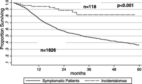
Methods: We reviewed 1944 consecutive pancreaticoduodenectomies (PD) over an 8-year period (April 1997 to October 2005). A total of 118 patients (6% of all PDs) presented with an incidental finding of a periampullary or pancreatic mass. The PI patients were analyzed and compared with the rest of the cohort (NI, nonincidentaloma group, n = 1826).
Results: Thirty-one percent of the PI patients (n = 37) had malignant disease (versus 76% of the NI patients, P < 0.001), 47% (n = 55) had premalignant disease, and the remaining 22% (n = 26) had little or no risk for malignant progression. The 3 most common diagnoses in the PI group were IPMN without invasive cancer (30%), cystadenoma (17%), and pancreatic ductal adenocarcinoma (10%). The PI group had a higher overall complication rate (55% versus 43%, P = 0.02), due in part to a significantly increased rate of pancreatic fistulas (18.4% PI versus 8.5% NI, P < 0.001). Patients in the PI group with malignant disease had a superior long-term survival (median, 30 months, P = 0.01) compared with patients in the NI group with malignant disease (median, 21 months).
Conclusions: Incidentally discovered periampullary and pancreatic masses comprise a substantial proportion of patients undergoing PD. Roughly three fourths of these lesions are malignant or premalignant, and amenable to curative resection. Resected malignant PIs have favorable pathologic features as compared with resected malignant NIs, and resection of these early lesions in asymptomatic individuals is associated with improved survival, compared with patients with symptomatic disease.
Figures


References
-
- emedicine CH. CT Scan Introduction. CT Scan. 2005 August 10, 2005 [cited 2005 October 14, 2005]; Available from: http://www.emedicinehealth.com/articles/11618–1.asp.
-
- Indrajit IK, D'souza JD. Multislice CT: a quantum leap in whole body imaging. Indian J Radiol Imaging. 2004 2004;14:209–216.
-
- Davis W. Multi-Slice CT: 64 and Counting. Med Imaging. 2005.
-
- Linton OW, Mettler FA Jr. National conference on dose reduction in CT, with an emphasis on pediatric patients. AJR Am J Roentgenol. 2003;181:321–329. - PubMed
-
- Brant-Zawadzki M. CT screening: why I do it. AJR Am J Roentgenol. 2002;179:319–326. - PubMed
MeSH terms
LinkOut - more resources
Full Text Sources
Medical
Research Materials
Miscellaneous
