Review
. 2006 Oct-Nov;26(7-8):1113-29.
doi: 10.1007/s10571-006-9007-2. Epub 2006 Apr 22.

Bone marrow stem cells and polymer hydrogels--two strategies for spinal cord injury repair

Affiliations
Review

Bone marrow stem cells and polymer hydrogels--two strategies for spinal cord injury repair

Eva Syková et al. Cell Mol Neurobiol. 2006 Oct-Nov.

Abstract

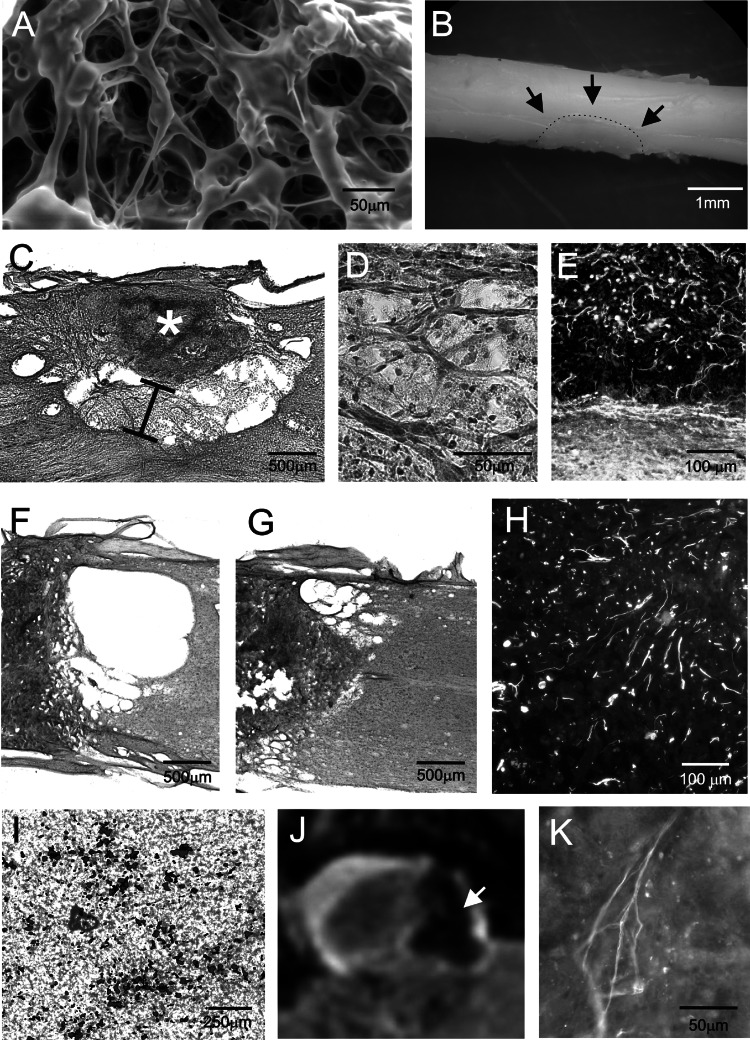
1. Emerging clinical studies of treating brain and spinal cord injury (SCI) led us to examine the effect of autologous adult stem cell transplantation as well as the use of polymer scaffolds in spinal cord regeneration. We compared an intravenous injection of mesenchymal stem cells (MSCs) or the injection of a freshly prepared mononuclear fraction of bone marrow cells (BMCs) on the treatment of an acute or chronic balloon-induced spinal cord compression lesion in rats. Based on our experimental studies, autologous BMC implantation has been used in a Phase I/II clinical trial in patients (n=20) with a transversal spinal cord lesion. 2. MSCs were isolated from rat bone marrow by their adherence to plastic, labeled with iron-oxide nanoparticles and expanded in vitro. Macroporous hydrogels based on derivatives of 2-hydroxyethyl methacrylate (HEMA) or 2-hydroxypropyl methacrylamide (HPMA) were prepared, then modified by their copolymerization with a hydrolytically degradable crosslinker, N,O-dimethacryloylhydroxylamine, or by different surface electric charges. Hydrogels or hydrogels seeded with MSCs were implanted into rats with hemisected spinal cords. 3. Lesioned animals grafted with MSCs or BMCs had smaller lesions 35 days postgrafting and higher scores in BBB testing than did control animals and also showed a faster recovery of sensitivity in their hind limbs using the plantar test. The functional improvement was more pronounced in MSC-treated rats. In MR images, the lesion populated by grafted cells appeared as a dark hypointense area and was considerably smaller than in control animals. Morphometric measurements showed an increase in the volume of spared white matter in cell-treated animals. In the clinical trial, we compared intraarterial (via a. vertebralis, n=6) versus intravenous administration of BMCs (n=14) in a group of subacute (10-33 days post-SCI, n=8) and chronic patients (2-18 months, n=12). For patient follow-up we used MEP, SEP, MRI, and the ASIA score. Our clinical study revealed that the implantation of BMCs into patients is safe, as there were no complications following cell administration. Partial improvement in the ASIA score and partial recovery of MEP or SEP have been observed in all subacute patients who received cells via a. vertebralis (n=4) and in one out of four subacute patients who received cells intravenously. Improvement was also found in one chronic patient who received cells via a. vertebralis. A much larger population of patients is needed before any conclusions can be drawn. The implantation of hydrogels into hemisected rat spinal cords showed that cellular ingrowth was most pronounced in copolymers of HEMA with a positive surface electric charge. Although most of the cells had the morphological properties of connective tissue elements, we found NF-160-positive axons invading all the implanted hydrogels from both the proximal and distal stumps. The biodegradable hydrogels degraded from the border that was in direct contact with the spinal cord tissue. They were resorbed by macrophages and replaced by newly formed tissue containing connective tissue elements, blood vessels, GFAP-positive astrocytic processes, and NF-160-positive neurofilaments. Additionally, we implanted hydrogels seeded with nanoparticle-labeled MSCs into hemisected rat spinal cords. Hydrogels seeded with MSCs were visible on MR images as hypointense areas, and subsequent Prussian blue histological staining confirmed positively stained cells within the hydrogels. 4. We conclude that treatment with different bone marrow cell populations had a positive effect on behavioral outcome and histopathological assessment after SCI in rats; this positive effect was most pronounced following MSC treatment. Our clinical study suggests a possible positive effect in patients with SCI. Bridging the lesion cavity can be an approach for further improving regeneration. Our preclinical studies showed that macroporous polymer hydrogels based on derivatives of HEMA or HPMA are suitable materials for bridging cavities after SCI; their chemical and physical properties can be modified to a specific use, and 3D implants seeded with different cell types may facilitate the ingrowth of axons.


Figures

Fig. 1.
Fig. 1.
Behavioural testing after treatment with MSCs and BMCs. A: Behavioural open field BBB motor scores of MSC- and BMC-treated rats were significantly higher than those of control animals 14, 21, 28, and 35 days after SCI (p < 0.05). B: Time course of the animals' response to radiant heat measured by the plantar test in treated and control rats. In all treated rats, latency times decreased as their recovery progressed. The most prominent effect was seen in MSC-treated rats. The latency time in saline-injected (control) rats did not change during the 35-day survival period. Data are averaged between right and left hind limbs and expressed as mean ± SEM. * p < 0.05 compared to control group.
Fig. 2.
Fig. 2.
MSCs labeled with nanoparticles implanted into rats with a spinal cord compression lesion. A,B: Transversal and longitudinal images of a spinal cord compression lesion populated by intravenously injected nanoparticle-labeled MSCs, four weeks after implantation. The lesion with nanoparticle-labeled cells is visible as a dark hypointensive area. C: Prussian blue staining of a spinal cord compression lesion (control animal). D: Prussian blue staining of a spinal cord lesion with intravenously injected nanoparticle-labeled MSCs. Note the smaller lesion size in the implanted animal than in the control. Insert: The lesion is populated with Pussian blue-positive cells. A–D Modified from (Jendelová et al., 2004b). E: Analysis of the spared white and grey matter volume in the centre of the lesion. The total volume of the white matter (WM) and grey matter (GM) in 11 mm long segments of the spinal lesion in treated and saline injected animals. No statistically significant differences in spared grey matter volume were observed between the groups. Data are represented as group means ± SEM. * p < 0.05 compared to control group.
Fig. 3.
Fig. 3.
The use of hydrogels in spinal cord injury repair. A: The structure of a HEMA hydrogel observed using a scanning electron microscope. B: Four weeks after implantation, a HEMA hydrogel firmly adheres to the spinal cord tissue. The dotted line marks the interface between spinal cord tissue and the hydrogel, which is fully integrated into the spinal cord. C: A biodegragable hydrogel (HPMA +10% EOEMA) degraded from the interface with the spinal cord towards the central part of the hydrogel. The size (volume) of the peripheral zone of degraded hydrogel is marked by the black line. The central part of the hydrogel (marked with an asterisk) consisted of amorphous matter, where mostly the ingrowth of connective tissue and capillaries was observed. D: The detail of the peripheral zone of a degraded hydrogel. The gel was resorbed by macrophages and replaced by newly formed tissue. E: The detail of the peripheral zone of a degraded hydrogel. NF160-positive neurofilaments are entering the gel from the spinal cord border. F: Interface between the spinal cord stump and a hydrogel acutely implanted into a complete spinal cord transection. G: Interface between the spinal cord stump and a hydrogel implanted seven days after complete spinal cord transection. Note the smaller cavity with delayed implantation. H: Immunostaining with anti-neurofilament antibody (anti-NF160) showing the presence of NF160-positive axons in the centre of a hydrogel inserted 7days after complete spinal cord transection. Images C–H were taken four weeks postimplantation. I: Endorem-labeled cells seeded into a hydrogel. J: A hydrogel seeded with MSCs was visible on MR images 6 weeks after implantation as a hypointensive area (arrow). K: Ingrowth of NF 160-positive neurofilaments into a hydrogel seeded with MSCs. I–J Modified from (Syková and Jendelová, 2005).

Similar articles

Cited by

References

    1. Akiyama, Y., Radtke, C., Honmou, O., and Kocsis, J. D. (2002b). Remyelination of the spinal cord following intravenous delivery of bone marrow cells. Glia39:229–236. - PMC - PubMed
    1. Akiyama, Y., Radtke, C., and Kocsis, J. D. (2002a). Remyelination of the rat spinal cord by transplantation of identified bone marrow stromal cells. J. Neurosci.22:6623–6630. - PMC - PubMed
    1. Azizi, S. A., Stokes, D., Augelli, B. J., DiGirolamo, C., and Prockop, D. J. (1998). Engraftment and migration of human bone marrow stromal cells implanted in the brains of albino rats–similarities to astrocyte grafts. Proc. Natl. Acad. Sci. U.S.A.95:3908–3913. - PMC - PubMed
    1. Basso, D. M., Beattie, M. S., Bresnahan, J. C., Anderson, D. K., Faden, A. I., Gruner, J. A., et al. (1996). MASCIS evaluation of open field locomotor scores: effects of experience and teamwork on reliability. Multicenter Animal Spinal Cord Injury Study. J. Neurotrauma13:343–359. - PubMed
    1. Bixby, J. L., and Harris, W. A. (1991). Molecular mechanisms of axon growth and guidance. Annu. Rev. Cell. Biol.7:117–159. - PubMed

Publication types

LinkOut - more resources