

Antimicrobial activities of silver used as a polymerization catalyst for a wound-healing matrix
- PMID: 16635526
- PMCID: PMC2077300
- DOI: 10.1016/j.biomaterials.2006.03.038
Antimicrobial activities of silver used as a polymerization catalyst for a wound-healing matrix
Abstract
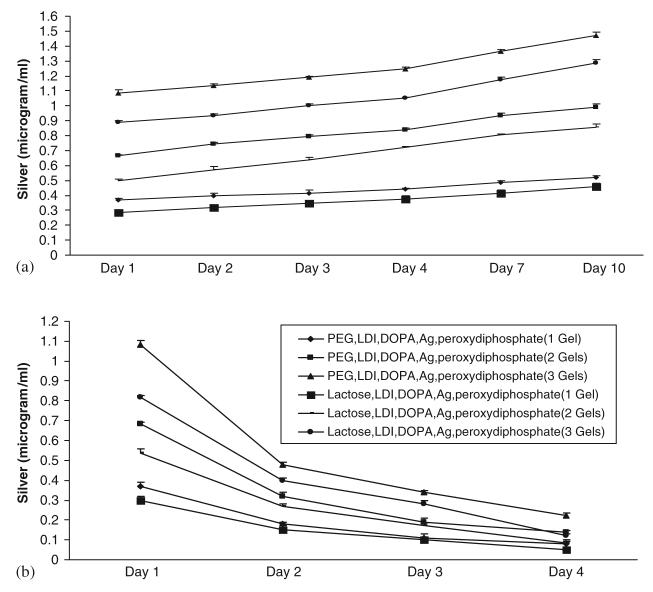
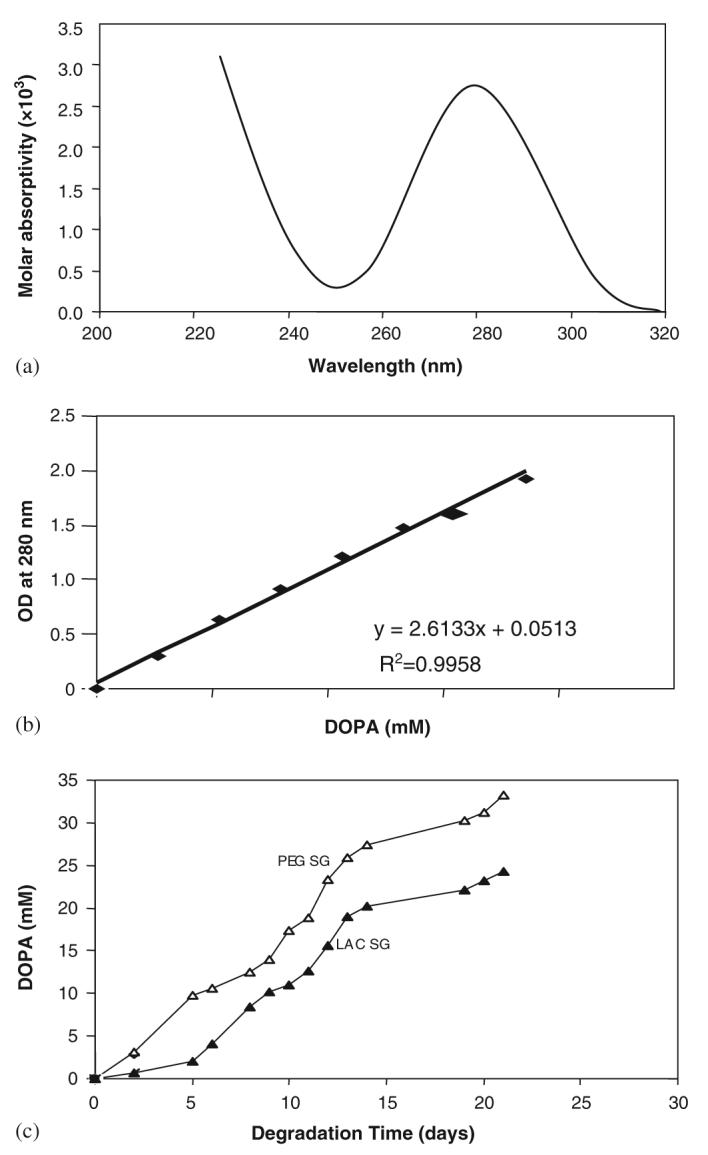
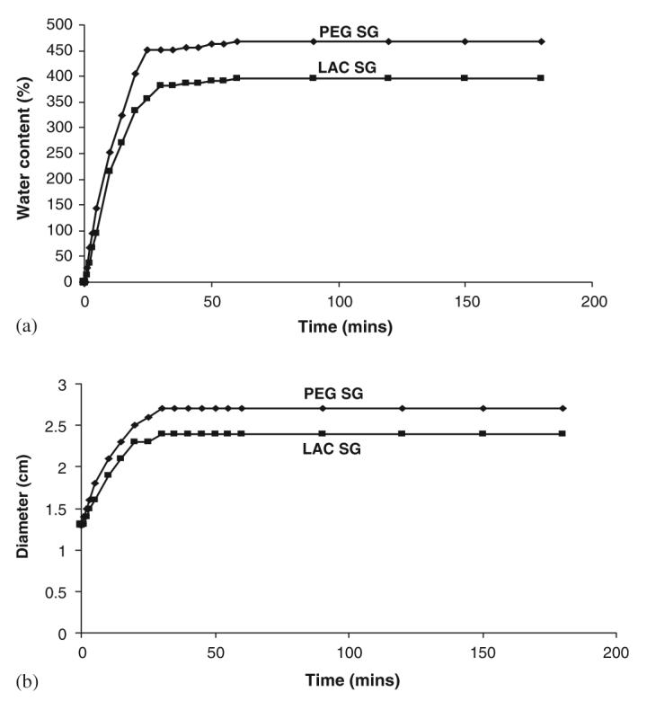
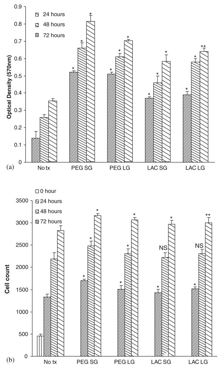
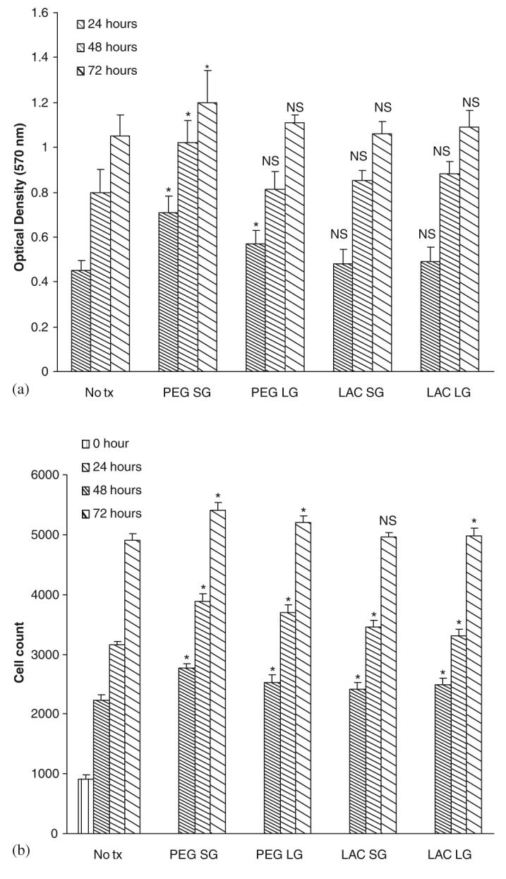
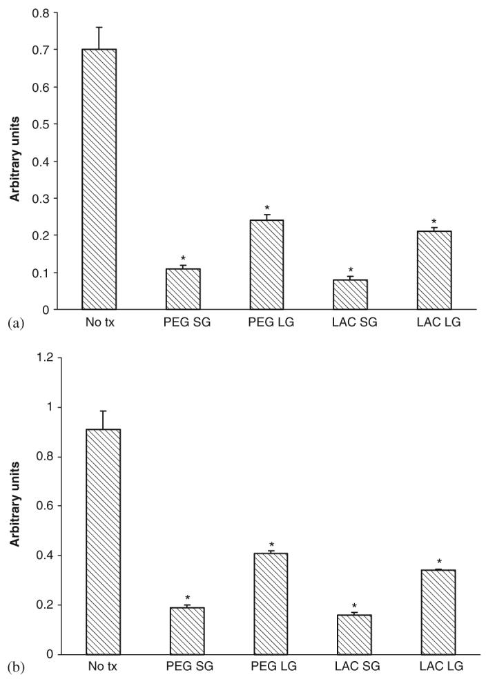
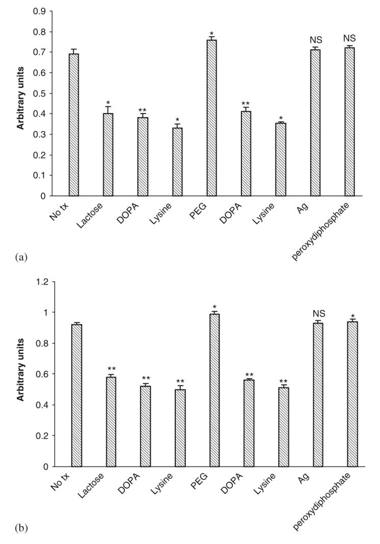
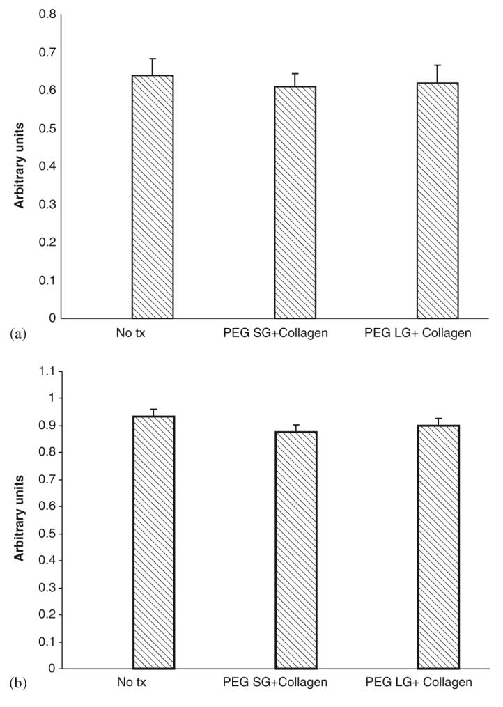
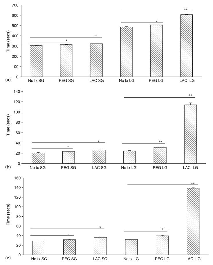
Wound healing is a complex and orchestrated process that re-establishes the barrier and other functions of the skin. While wound healing proceeds apace in healthy individual, bacterial overgrowth and infection disrupts this process with significant morbidity and mortality. As such, any artificial matrix to promote wound healing must also control infecting microbes. We had earlier developed a two-part space-conforming gel backbone based on polyethyleneglycol (PEG) or lactose, which used ionic silver as the catalyst for gelation. As silver is widely used as an in vitro antimicrobial, use of silver as a catalyst for gelation provided the opportunity to assess its function as an anti-microbial agent in the gels. We found that these gels show bacteriostatic and bactericidal activity for a range of Gram-negative and Gram-positive organisms, including aerobic as well as anaerobic bacteria. This activity lasted for days, as silver leached out of the formed gels over a day in the manner of second-order decay. Importantly the gels did not limit either cell growth or viability, though cell migration was affected. Adding collagen I fragments to the gels corrected this effect on cell migration. We also found that the PEG gel did not interfere with hemostasis. These observations provide the basis for use of the gel backbones for incorporation of anesthetic agents and factors that promote wound repair. In conclusion, silver ions can serve dual functions of catalyzing gelation and providing anti-microbial properties to a biocompatible polymer.
Figures
Similar articles
-
Silver nanoparticle impregnated chitosan-PEG hydrogel enhances wound healing in diabetes induced rabbits.Int J Pharm. 2019 Mar 25;559:23-36. doi: 10.1016/j.ijpharm.2019.01.019. Epub 2019 Jan 19. Int J Pharm. 2019. PMID: 30668991
-
Silver nanoparticles in therapeutics: development of an antimicrobial gel formulation for topical use.Mol Pharm. 2009 Sep-Oct;6(5):1388-401. doi: 10.1021/mp900056g. Mol Pharm. 2009. PMID: 19473014
-
Infection-Free and Enhanced Wound Healing Potential of Alginate Gels Incorporating Silver and Tannylated Calcium Peroxide Nanoparticles.Int J Mol Sci. 2024 May 10;25(10):5196. doi: 10.3390/ijms25105196. Int J Mol Sci. 2024. PMID: 38791232 Free PMC article.
-
Silver-based gels for oral and skin infections: antimicrobial effect and physicochemical stability.Future Microbiol. 2023 Sep;18:985-996. doi: 10.2217/fmb-2023-0034. Epub 2023 Sep 26. Future Microbiol. 2023. PMID: 37750752 Review.
-
The growing importance of materials that prevent microbial adhesion: antimicrobial effect of medical devices containing silver.Int J Antimicrob Agents. 2009 Aug;34(2):103-10. doi: 10.1016/j.ijantimicag.2009.01.017. Epub 2009 Mar 31. Int J Antimicrob Agents. 2009. PMID: 19339161 Review.
Cited by
-
The effect of multifunctional polymer-based gels on wound healing in full thickness bacteria-contaminated mouse skin wound models.Biomaterials. 2007 Sep;28(27):3977-86. doi: 10.1016/j.biomaterials.2007.05.008. Epub 2007 May 24. Biomaterials. 2007. PMID: 17561250 Free PMC article.
-
Structural and physical properties of antibacterial Ag-doped nano-hydroxyapatite synthesized at 100°C.Nanoscale Res Lett. 2011 Dec 3;6(1):613. doi: 10.1186/1556-276X-6-613. Nanoscale Res Lett. 2011. PMID: 22136671 Free PMC article.
-
Sex affects the response of Wistar rats to polyvinyl pyrrolidone (PVP)-coated silver nanoparticles in an oral 28 days repeated dose toxicity study.Part Fibre Toxicol. 2021 Oct 18;18(1):38. doi: 10.1186/s12989-021-00425-y. Part Fibre Toxicol. 2021. PMID: 34663357 Free PMC article.
-
Improved Transplanted Stem Cell Survival in a Polymer Gel Supplemented With Tenascin C Accelerates Healing and Reduces Scarring of Murine Skin Wounds.Cell Transplant. 2017 Jan 24;26(1):103-113. doi: 10.3727/096368916X692249. Epub 2016 Jul 22. Cell Transplant. 2017. PMID: 27452449 Free PMC article.
-
Preparation and properties of nano-sized Ag and Ag2S particles in biopolymer matrix.Eur Phys J E Soft Matter. 2007 Jan;22(1):51-9. doi: 10.1140/epje/e2007-00008-y. Epub 2007 Feb 21. Eur Phys J E Soft Matter. 2007. PMID: 17318289
References
-
- Babu R, Zhang JY, Beckman E, Pasculle AW, Wells A. A polymer-based gel to promote wound healing. Am J Clin Pathol. 2005;124(3):455.
-
- Amadeu TP, Coulomb B, Desmouliere A, Costa AM. Cutaneous wound healing: myofibroblastic differentiation and in vitro models. Int J Low Extrem Wounds. 2003;2(2):60–8. - PubMed
-
- Klein DG, Fritsch DE, Amin SG. Wound infection following trauma and burn injuries. Crit Care Nurs Clin North Am. 1995;7(4):627–42. - PubMed
-
- Ovington L. Bacterial toxins and wound healing. Ostomy Wound Manage. 2003;49(7A suppl):8–12. - PubMed
-
- Guggenbicher JP, Boswald M, Lugauer S, Krall T. A new technology of microdispersed silver in polyurethane induces antimicrobial activity in central venous catheters. Infection. 1999;27(Suppl 1):S16–23. - PubMed
Publication types
MeSH terms
Substances
Grants and funding
LinkOut - more resources
Full Text Sources
Other Literature Sources
Medical
Research Materials
