. 2006 Apr 24;173(2):231-9.
doi: 10.1083/jcb.200511103.

Lysosomal biogenesis and function is critical for necrotic cell death in Caenorhabditis elegans

Affiliations

Lysosomal biogenesis and function is critical for necrotic cell death in Caenorhabditis elegans

Marta Artal-Sanz et al. J Cell Biol. .

Abstract

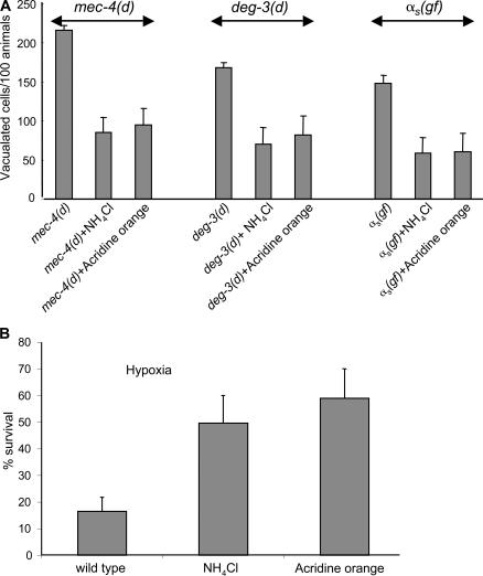
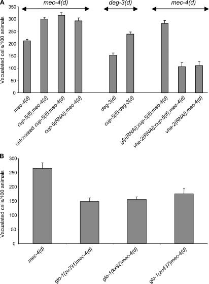
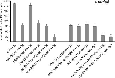
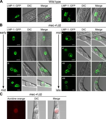
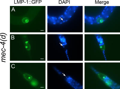
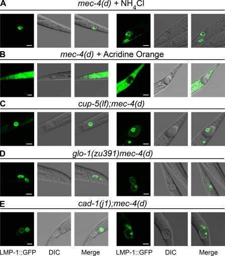
Necrotic cell death is defined by distinctive morphological characteristics that are displayed by dying cells (Walker, N.I., B.V. Harmon, G.C. Gobe, and J.F. Kerr. 1988. Methods Achiev. Exp. Pathol. 13:18-54). The cellular events that transpire during necrosis to generate these necrotic traits are poorly understood. Recent studies in the nematode Caenorhabditis elegans show that cytoplasmic acidification develops during necrosis and is required for cell death (Syntichaki, P., C. Samara, and N. Tavernarakis. 2005. Curr. Biol. 15:1249-1254). However, the origin of cytoplasmic acidification remains elusive. We show that the alkalization of endosomal and lysosomal compartments ameliorates necrotic cell death triggered by diverse stimuli. In addition, mutations in genes that result in altered lysosomal biogenesis and function markedly affect neuronal necrosis. We used a genetically encoded fluorescent marker to follow lysosome fate during neurodegeneration in vivo. Strikingly, we found that lysosomes fuse and localize exclusively around a swollen nucleus. In the advanced stages of cell death, the nucleus condenses and migrates toward the periphery of the cell, whereas green fluorescent protein-labeled lysosomal membranes fade, indicating lysosomal rupture. Our findings demonstrate a prominent role for lysosomes in cellular destruction during necrotic cell death, which is likely conserved in metazoans.


Figures

Figure 1.
Figure 1.
Alkalization of endosomal compartments by weak bases protects against necrotic cell death. (A) Degenerating touch receptor neurons in mec-4(d) and PVC interneurons in deg-3(d) and αs (gf) in untreated animals or animals treated with NH4Cl and acridine orange. n > 150. P < 0.001, unpaired t test. (B) The effects of alkalizing agents on hypoxic death. The graph shows the percentage of animals that survived near-lethal treatment with sodium azide. n > 200. P < 0.001; unpaired t test. Error bars represent the SD of the mean.
Figure 2.
Figure 2.
Effect of lysosomal biogenesis mutants on necrotic cell death. (A) Neurodegeneration is enhanced in a cup-5(lf) genetic background, where the lysosomal system is expanded. This enhancement is suppressed by vacuolar H+-ATPase deficiency. Degenerating neurons in mec-4(d), cup-5(lf);mec-4(d), outcrossed cup-5(lf);mec-4(d), cup-5(RNAi);mec-4(d), deg-3(d), and cup-5(lf);deg-3(d) animals. Knockdown of cup-5 by RNAi also enhances mec-4(d)–induced neurodegeneration. n > 250. P < 0.005; unpaired t test. (B) Neurodegeneration is suppressed by three different glo-1 mutant alleles, where lysosomal biogenesis is defective. Degenerating neurons in mec-4(d), glo-1(zu391)mec-4(d), glo-1(kx92)mec-4(d), and glo-1(zu437)mec-4(d) animals. n > 350. P < 0.005, unpaired t test. Error bars represent the SD of the mean.
Figure 3.
Figure 3.
Suppression of necrosis by aspartyl protease deficiency is enhanced by conditions that impede intracellular acidification. The number of vacuolated touch receptor neurons per 100 L1 stage mec-4(d) animals. Bars represent the mean of three independent experiments. n > 300. Knockdown of both vacuolar H+-ATPase and aspartyl protease genes resulted in significantly more extended quenching of neurodegeneration than for any single gene. P < 0.001, unpaired t test. Efficacy of RNAi was assessed as described in Materials and methods. Error bars represent the SD of the mean.
Figure 4.
Figure 4.
Lysosomal morphology and distribution during neurodegeneration. (A) Confocal images of wild-type touch receptor neurons expressing a pmec-17 LMP-1∷GFP transgene. LMP-1∷GFP expression, differential interference contrast (DIC), and merged images are shown. Healthy neurons show a scattered and punctate pattern of lysosomal distribution. (i) Wild-type PVM touch receptor neuron. (ii) Wild-type ALM (left and right) touch receptor neurons. (B) Confocal images of PLM touch receptor neurons of mec-4(d) animals expressing a pmec-17LMP-1∷GFP transgene. During the early to middle (mid) stages of degeneration, lysosomes enlarge and appear to coalesce around a swollen nucleus (i–iv). Later on, the nucleus migrates to the periphery of the cell and condenses (iv–vi). At the late stage, no lysosome structure is evident and the vacuolated cell becomes diffusely fluorescent (vii and viii). (C) Acridine orange staining of a middle to late degenerating PLM touch receptor neuron. Acridine orange, DIC, and merged images are shown. Bars, 5 μm.
Figure 5.
Figure 5.
DAPI staining of mec-4(d) animals expressing pmec-17 LMP-1∷GFP. (A) ALM touch receptor neuron. (B and C) PLM touch receptor neurons. Arrows point to DAPI-positive nuclei of the dying neurons. Bars, 5 μm.
Figure 6.
Figure 6.
Perturbation of lysosomal biogenesis, function, and neurodegeneration. Lysosomal morphology and distribution after treatment with alkalizing agents and in genetic backgrounds affecting lysosomal biogenesis and function. (A and B) Confocal images of PLM touch receptor neurons of mec-4(d) animals expressing a pmec-17LMP-1∷GFP reporter fusion, after alkalization of lysosomal compartments. (A) Images of degenerating neurons after treatment with 5 mM NH4Cl. (B) Degenerating neurons after treatment with 40 μM acridine orange. Nuclei can be seen because, at the concentration used, acridine orange stains DNA. (C–E) Confocal images of PLM touch receptor neurons of mec-4(d) animals under different genetic backgrounds that either suppress or enhance necrosis. Double mutants express the same pmec-17LMP-1∷GFP transgene. (C) Degenerating neurons of cup-5(lf);mec-4(d) mutants. Note that vacuoles appear larger. (D) Degenerating neurons of glo-1(zu391) mec-4(d) animals. (E) Images of degenerating neurons in cad-1(j1);mec-4(d) mutants. LMP-1∷GFP expression, differential interference contrast (DIC), and merged images are shown. (left) Early to middle stages of neurodegeneration. (right) Middle to late stages of neurodegeneration. Bars, 5 μm.

Similar articles

Cited by

References

    1. Artal-Sanz, M., and N. Tavernarakis. 2005. Proteolytic mechanisms in necrotic cell death and neurodegeneration. FEBS Lett. 579:3287–3296. - PubMed
    1. Bakker, A.C., P. Webster, W.A. Jacob, and N.W. Andrews. 1997. Homotypic fusion between aggregated lysosomes triggered by elevated [Ca2+]i in fibroblasts. J. Cell Sci. 110:2227–2238. - PubMed
    1. Brenner, S. 1974. The genetics of Caenorhabditis elegans. Genetics. 77:71–94. - PMC - PubMed
    1. Burkhardt, J.K., C.J. Echeverri, T. Nilsson, and R.B. Vallee. 1997. Overexpression of the dynamitin (p50) subunit of the dynactin complex disrupts dynein-dependent maintenance of membrane organelle distribution. J. Cell Biol. 139:469–484. - PMC - PubMed
    1. Carafoli, E. 2002. Calcium signaling: a tale for all seasons. Proc. Natl. Acad. Sci. USA. 99:1115–1122. - PMC - PubMed

Publication types