. 2006 May 2;103(18):7148-53.
doi: 10.1073/pnas.0602048103. Epub 2006 Apr 24.

Disulfide cross-linked protein represents a significant fraction of ALS-associated Cu, Zn-superoxide dismutase aggregates in spinal cords of model mice

Affiliations

Disulfide cross-linked protein represents a significant fraction of ALS-associated Cu, Zn-superoxide dismutase aggregates in spinal cords of model mice

Yoshiaki Furukawa et al. Proc Natl Acad Sci U S A. .

Abstract

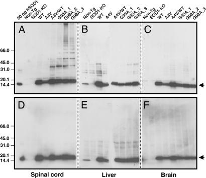
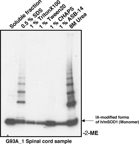
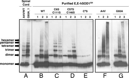
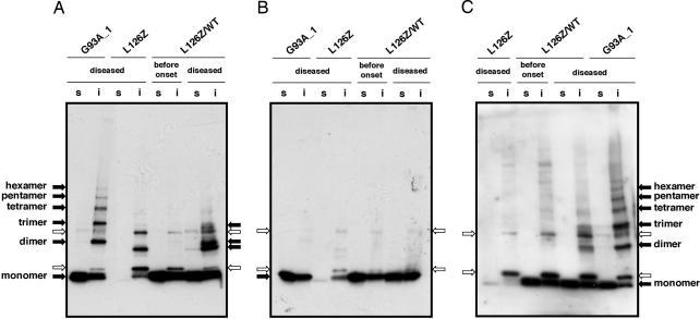
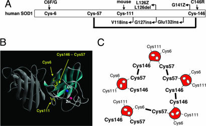
Point mutations in Cu, Zn-superoxide dismutase (SOD1) cause a familial form of the neurodegenerative disease amyotrophic lateral sclerosis (ALS). Aggregates of mutant SOD1 proteins are observed in histopathology and are invoked in several proposed mechanisms for motor neuronal death; however, the significant stability and activity of the mature mutant proteins are not readily explained in such models. Recent biochemical studies suggest that it is the immature disulfide-reduced forms of the familial ALS mutant SOD1 proteins that play a critical role; these forms tend to misfold, oligomerize, and readily undergo incorrect disulfide formation upon mild oxidative stress in vitro. Here we provide physiological support for this mechanism of aggregate formation and show that a significant fraction of the insoluble SOD1 aggregates in spinal cord of the ALS-model transgenic mice contain multimers cross-linked via intermolecular disulfide bonds. These insoluble disulfide-linked SOD1 multimers are found only in the spinal cord of symptomatic transgenic animals, are not observed in unafflicted tissue such as brain cortex and liver, and can incorporate WT SOD1 protein. The findings provide a biochemical basis for a pathological hallmark of this disease; namely, incorrect disulfide cross-linking of the immature, misfolded mutant proteins leads to insoluble aggregates.


Conflict of interest statement

Conflict of interest statement: No conflicts declared.

Figures

Fig. 1.
Fig. 1.
SOD1 disulfide-linked multimers found in the mouse tissue samples. Each tissue sample, i.e., spinal cord (A and D), liver (B and E), and brain (C and F), was ground in the presence of 100 mM IA and 2.5% SDS to protect the free thiol groups from the oxidation during isolation and electrophoresis. Six micrograms of total proteins was loaded on 4–15% SDS/PAGE gradient gel without (AC) or with (DF) 2-ME, and the SOD1 proteins were detected by using Western blot. As a positive control, 50 ng of human Cu, Zn-superoxide dismutase (Sigma) also was loaded. Arrows indicate IA-modified forms of h/mSOD1 (monomer).
Fig. 2.
Fig. 2.
Solubility test of SOD1 disulfide-linked multimers. Spinal cord tissue of the G93A transgenic mouse was ground in the presence of 100 mM IA but in the absence of any detergents. After incubation at 37°C for 1 h, the supernatant was obtained by centrifugation and used as the soluble fraction. The protein pellet was redissolved in the buffer containing 0.5% SDS, 1% Triton X-100, 1% Tween 20, 1% CHAPS, 1% ASB-14, or 8 M urea. Each supernatant after centrifugation (6 μg of total proteins) was loaded on 4–15% SDS/PAGE gradient gel, and protein bands are developed with Western blot.
Fig. 3.
Fig. 3.
SOD1 disulfide-linked multimers derived from purified proteins. In anaerobic conditions, a 3 μM concentration of E,E-SOD1SH proteins [WT (B), C6S/C111S (C), C57S/C146S (D), C4S (E), A4V (F), and G93A (G)] were incubated for 1 h with 50 mM DTT, 2 mM GSH/4 mM GSSG, or 10 mM H2O2 in 50 mM Hepes/0.1 mM EDTA//0.1 mM BCS, pH 7.2. Before being loaded onto 4–15% SDS/PAGE gradient gel, free thiol groups were protected by reaction with 100 mM IA in the presence of 2.5% SDS. Lanes: 1, 5 mM DTT; 2, 2 mM GSH/4 mM GSSG; 3, 10 mM H2O2. One hundred nanograms of proteins was analyzed by Western blot with SOD1 polyclonal antibody. For comparison, insoluble fractions (6 μg of total proteins) of the A4V/WT and G93A spinal cord tissues (A) that are dissolved in 0.5% SDS also are shown at the left side of the figure.
Fig. 4.
Fig. 4.
Disulfide-linked SOD1 dimerization in the diseased mouse spinal cord expressing truncated protein, L126Z. The spinal cord tissues of G93A, L126Z, and L126Z/WT are ground in the presence of 100 mM IA but in the absence of any detergents. After incubation at 37°C for 1 h, the supernatant (s, soluble fraction) was obtained by centrifugation. The protein pellet was redissolved in the buffer containing 100 mM IA and 0.5% SDS, and the supernatant after centrifugation was used as an insoluble fraction (i). (A and B) The polyclonal antibody to SOD1 from EMD Biosciences or the polyclonal antibody to C-terminal region of SOD1 (C) was used. 2-ME is included in the sample loading buffer in B but not in A and C. Nonspecific and SOD1-specific protein bands are indicated by white and black arrows, respectively (see Fig. 7). For analysis of soluble fractions, 6 μg of total proteins was used. Twelve micrograms of total proteins was analyzed in the insoluble fractions of L126Z and L126Z/WT, whereas 6 μg was used for analysis of the G93A insoluble fractions.
Fig. 5.
Fig. 5.
Importance of SOD1 Cys residues in ALS-associated mutations and disulfide-linked protein aggregation. (A) Cys-6 and -146 have been shown to link with fALS. Several truncated mutations in SOD1 lack Cys-146. Mouse SOD1 does not have Cys residue at position 111 but has serine. (B) Crystal structure of holoform of human SOD1 (1HL5). In the active form of SOD1, Cys-57 and -146 form the intramolecular disulfide bond, whereas Cys-6 and -111 are intact. (C) Cys-57 and -146 exhibit higher reactivity to form disulfide compared with Cys-6 and -111 (Fig. 3). Although Cys-146 is necessary for extensive disulfide-linked multimerization (Fig. 4), several pairs of Cys residues, i.e., Cys-57-Cys-57, Cys-57-Cys-146, and Cys-146-Cys-146, are possible. Note, nonconserved Cys residues also may contribute to the formation of disulfide-linked multimers.

Similar articles

Cited by

References

    1. Rosen D. R., Siddique T., Patterson D., Figlewicz D. A., Sapp P., Hentati A., Donaldson D., Goto J., O'Regan J. P., Deng H. X., et al. Nature. 1993;362:59–62. - PubMed
    1. Deng H. X., Hentati A., Tainer J. A., Iqbal Z., Cayabyab A., Hung W. Y., Getzoff E. D., Hu P., Herzfeldt B., Roos R. P., et al. Science. 1993;261:1047–1051. - PubMed
    1. McCord J. M., Fridovich I. J. Biol. Chem. 1969;244:6049–6055. - PubMed
    1. Reaume A. G., Elliott J. L., Hoffman E. K., Kowall N. W., Ferrante R. J., Siwek D. F., Wilcox H. M., Flood D. G., Beal M. F., Brown R. H., Jr, et al. Nat. Genet. 1996;13:43–47. - PubMed
    1. Liochev S. I., Fridovich I. Free Radical Biol. Med. 2003;34:1383–1389. - PubMed

Publication types