. 2006 May 2;103(18):7124-9.
doi: 10.1073/pnas.0510198103. Epub 2006 Apr 24.

Sleep after spatial learning promotes covert reorganization of brain activity

Affiliations

Sleep after spatial learning promotes covert reorganization of brain activity

Pierre Orban et al. Proc Natl Acad Sci U S A. .

Abstract

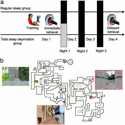
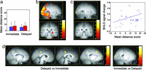
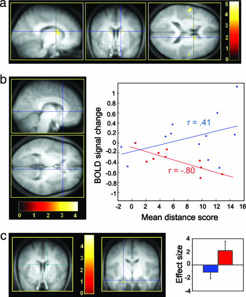
Sleep promotes the integration of recently acquired spatial memories into cerebral networks for the long term. In this study, we examined how sleep deprivation hinders this consolidation process. Using functional MRI, we mapped regional cerebral activity during place-finding navigation in a virtual town, immediately after learning and 3 days later, in subjects either allowed regular sleep (RS) or totally sleep-deprived (TSD) on the first posttraining night. At immediate and delayed retrieval, place-finding navigation elicited increased brain activity in an extended hippocampo-neocortical network in both RS and TSD subjects. Behavioral performance was equivalent between groups. However, striatal navigation-related activity increased more at delayed retrieval in RS than in TSD subjects. Furthermore, correlations between striatal response and behavioral performance, as well as functional connectivity between the striatum and the hippocampus, were modulated by posttraining sleep. These data suggest that brain activity is restructured during sleep in such a way that navigation in the virtual environment, initially related to a hippocampus-dependent spatial strategy, becomes progressively contingent in part on a response-based strategy mediated by the striatum. Both neural strategies eventually relate to equivalent performance levels, indicating that covert reorganization of brain patterns underlying navigation after sleep is not necessarily accompanied by overt changes in behavior.


Conflict of interest statement

Conflict of interest statement: No conflicts declared.

Figures

Fig. 1.
Fig. 1.
Experimental protocol and memory task. (a) Experimental design. After training on a desktop computer and subsequent testing in the scanner on day 1 (immediate retrieval), subjects were either TSD or allowed RS on the first posttraining night. They were all retested under identical conditions during a second fMRI session on day 4 (delayed retrieval). (b) The map depicts an aerial view of the color 3D virtual town in which subjects navigated at the ground level. Snapshots show the three locations used as targets for testing during the fMRI sessions. The 10 starting points are represented by numbers, with associated symbols indicating the target location to reach.
Fig. 2.
Fig. 2.
Navigation accuracy and place-finding-related brain network. Contrasts are displayed at P < 0.001 (uncorrected) superimposed on the average T1-weighted MRI scan. Color bars code the value of the t statistic associated with each voxel. (a) Mean distance scores at the immediate (on the left) and delayed (on the right) retrieval sessions for the RS (blue) and TSD (red) groups. Error bars are standard deviations. (b) Hippocampo-neocortical network activated in both populations during navigation in the virtual town at immediate retrieval (sagittal and coronal sections). Blue crosshair, right hippocampus (30, −30, 0 mm; Z = 4.33). (c) Brain areas whose activity positively correlated, at the between-subjects level, with accuracy of place finding at immediate retrieval (sagittal and coronal sections). Blue crosshair, right hippocampus (30, −32, 2 mm; Z = 3.65). The scatter plot shows the positive correlation (r = 0.32) between each subject's overall performance and level of activity in the right hippocampus, at the same coordinates. (d) Changes in place-finding-related brain activity between immediate and delayed retrieval sessions (coronal sections). (Left) Brain areas more activated at delayed than immediate retrieval. Blue crosshair, left caudate nucleus (−14, 20, 18 mm; Z = 3.57), right middle cingulate cortex (4, −34, 50 mm; Z = 3.81), right precuneus (2, −62, 50 mm; Z = 3.34), and right dorsolateral prefrontal cortex (18, 58, 18 mm; Z = 4.31). (Right) Decreased activity in the hippocampal complex at delayed as compared with immediate retrieval. Blue crosshair, left hippocampal area (−20, −28, −18 mm; Z = 4.40).
Fig. 3.
Fig. 3.
Sleep-dependent modulation of brain activity. Contrasts are displayed at P < 0.001 (uncorrected) superimposed on the average T1-weighted MRI scan. Color bars code the value of the t statistic associated with each voxel. (a) Higher activity elicited by place finding for the RS compared with the TSD group at delayed retrieval bilaterally in the caudate nucleus (sagittal, coronal, and axial sections). Blue crosshair, right caudate nucleus (14, 8, 18 mm; Z = 3.73). (b) Between-group regression analyses of the average session performance on cerebral activity at delayed retrieval (sagittal and coronal sections). Blue crosshair, right caudate nucleus (8, 22, 4 mm; Z = 3.45). The scatter plot shows that brain response at this coordinate was correlated positively with performance in the RS group (blue; r = 0.41) but negatively in the TSD group (red; r = −0.80). (c) Psychophysiological interaction analysis using the right caudate nucleus (14, 8, 18 mm; green crosshair) as seed area. The coupling of activity between the caudate nucleus and the left hippocampus (coronal section) was negative in the RS group (blue) but positive in the TSD group (red). Blue crosshair, left hippocampus (22, 12, 22 mm; Z = 3.15). Blue and red plots show the size of effect for each group. Error bars are standard deviations.

References

    1. Maquet P., Smith C., Stickgold R. Sleep and Brain Plasticity. Oxford: Oxford Univ. Press; 2003.
    1. Peigneux P., Laureys S., Delbeuck X., Maquet P. NeuroReport. 2001;12:A111–A124. - PubMed
    1. Gais S., Born J. Learn. Mem. 2004;11:679–685. - PMC - PubMed
    1. Rauchs G., Desgranges B., Foret J., Eustache F. J. Sleep Res. 2005;14:123–140. - PubMed
    1. Walker M. P., Stickgold R. Annu. Rev. Psychol. 2006;57:1–28. - PubMed

Publication types