. 2006 May;231(5):619-31.
doi: 10.1177/153537020623100517.

Retinoic acid exerts dual regulatory actions on the expression and nuclear localization of interferon regulatory factor-1

Affiliations

Retinoic acid exerts dual regulatory actions on the expression and nuclear localization of interferon regulatory factor-1

Xin M Luo et al. Exp Biol Med (Maywood). 2006 May.

Abstract

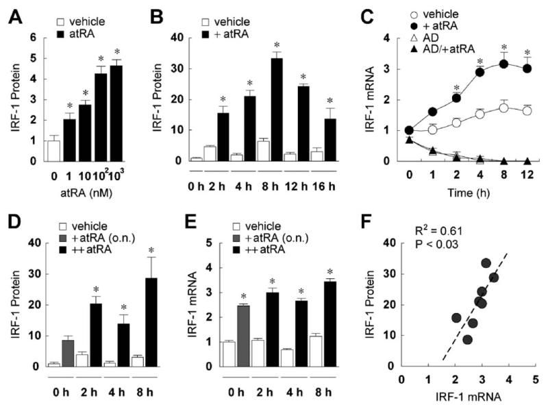
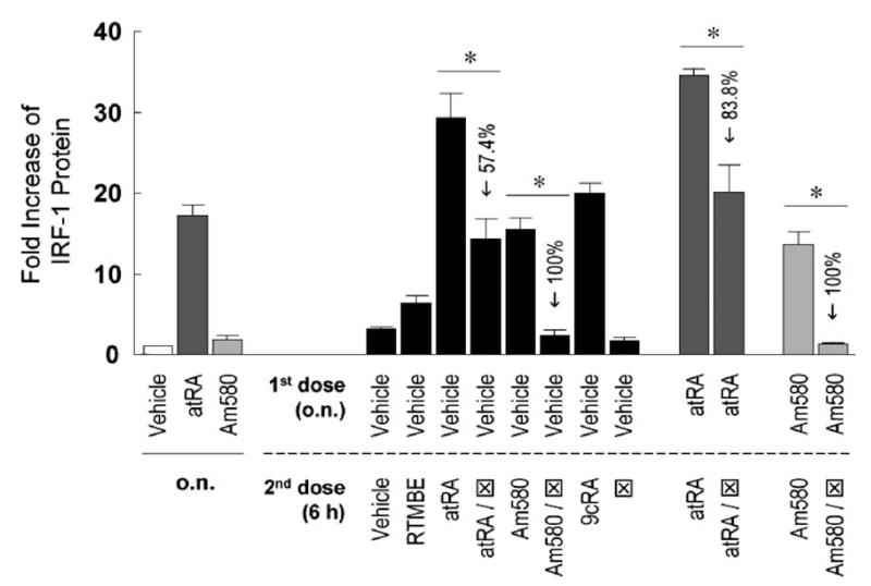
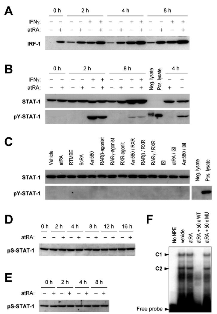
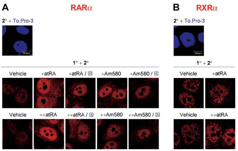
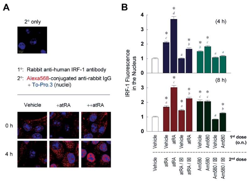
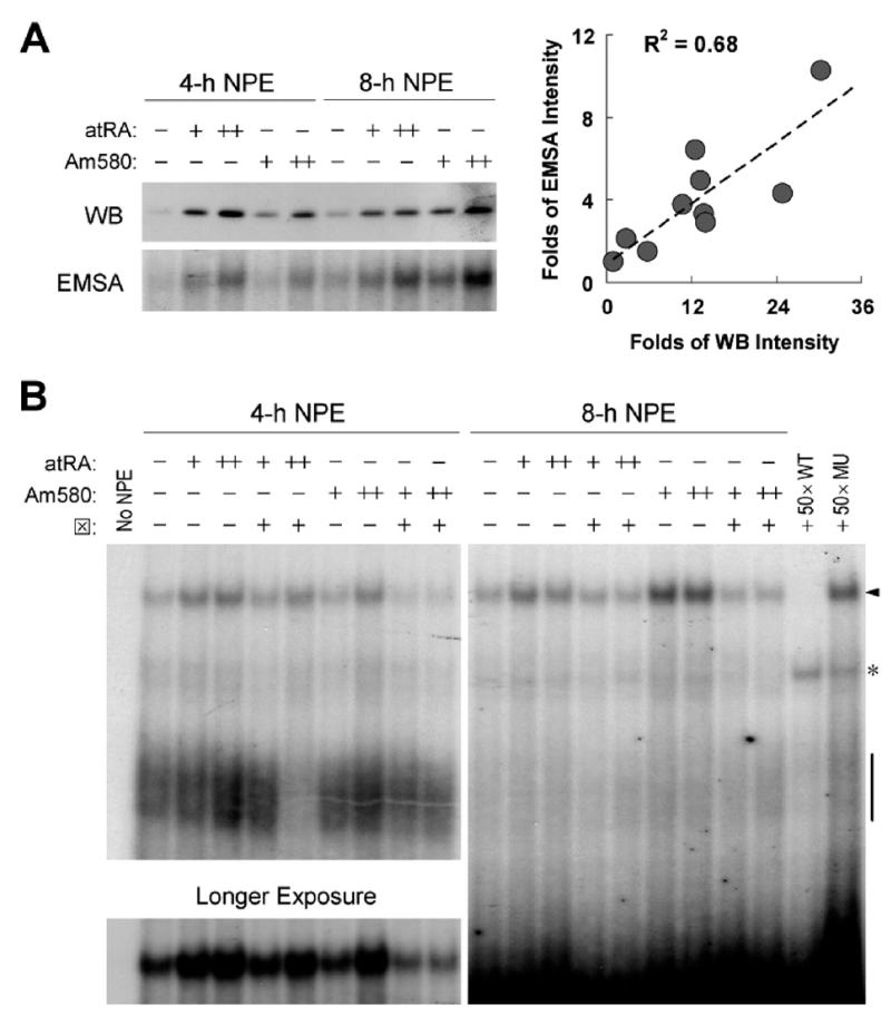
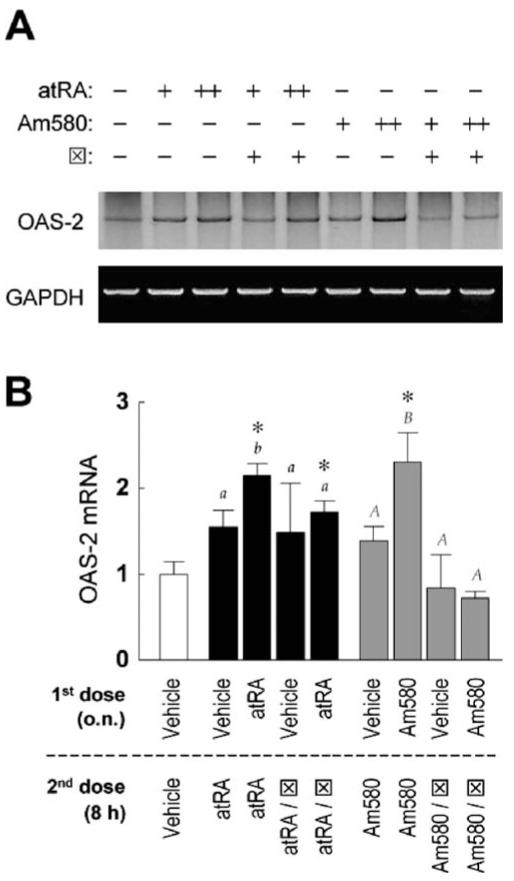
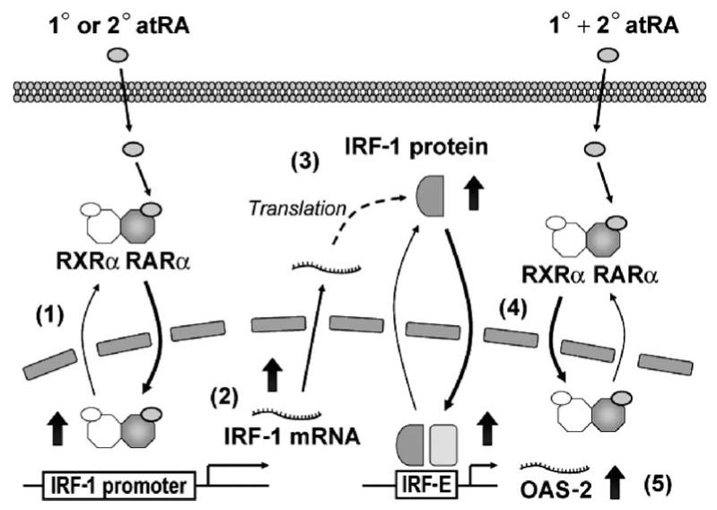
Interferon regulatory factor-1 (IRF-1), a transcription factor and tumor suppressor involved in cell growth regulation and immune responses, has been shown to be induced by all-trans retinoic acid (ATRA). However, the factors controlling the cellular location and activity of IRF-1 are not well understood. In this study, we examined the expression of IRF-1 and its nuclear localization, DNA-binding activity, and target gene expression in human mammary epithelial MCF10A cells, a model of breast epithelial cell differentiation and carcinogenesis. Following initial treatment with ATRA, IRF-1 mRNA and protein were induced within 2 hrs, reached a peak (>30-fold induction) at 8 hrs, and declined afterwards. IRF-1 protein was predominantly cytoplasmic during this treatment. Although a second dose of ATRA or Am580 (a related retinoid selective for retinoic acid receptor-alpha [RARalpha]), given 16 hrs after the first dose, restimulated IRF-1 mRNA and protein levels to a similar level to that obtained by the first dose, IRF-1 was predominantly concentrated in the nucleus after restimulation. ATRA and Am580 also increased nuclear RARalpha, whereas retinoid X receptor-alpha (RXRalpha)--a dimerization partner for RARalpha, was localized to the nucleus upon second exposure to ATRA. However, ATRA and Am580 did not regulate the expression or activation of signal transducer and activator of transcription-1 (STAT-1), a transcription factor capable of inducing the expression of IRF-1, indicating an STAT-1-independent mechanism of regulation by ATRA and Am580. The increase in nuclear IRF-1 after retinoid restimulation was accompanied by enhanced binding to an IRF-E DNA response element, and elevated expression of an IRF-1 target gene, 2',5'-oligoadenylate synthetase-2. The dual effect of retinoids in increasing IRF-1 mRNA and protein and in augmenting the nuclear localization of IRF-1 protein may be essential for maximizing the tumor suppressor activity and the immunosurveillance functions of IRF-1 in breast epithelial cells.


Figures

Figure 1
Figure 1
ATRA induces IRF-1 in MCF10A cells. (A) MCF10A cells were treated with vehicle or ATRA (1, 10, 100, or 1000 nM) overnight, and then lysed and assayed by immunoblotting for IRF-1 expression. The membranes were also blotted for β-actin to assure equal loading of proteins (data not shown). Fold increases of IRF-1 protein over the vehicle-treated level are shown (n = 3). ATRA-treated cells had significantly higher levels of IRF-1 protein as determined by one-way ANOVA (*). (B) Cells were treated with vehicle or ATRA (0.1 μM), cultured for 2–16 hrs, and then lysed and assayed by immunoblotting. Fold increases of IRF-1 protein compared to the untreated level at time 0 hrs are shown (n = 3). Two-way ANOVA showed ATRA and time as significant factors. For all time points, +ATRA was significantly different from vehicle (*). (C)Cells were treated with vehicle or ATRA (0.1 μM) for 1–12 hrs and RT-PCR was performed to assess the mRNA levels of IRF-1 and GAPDH. In some experiments, cells were pretreated with AD (5 μg/ml) 1 hr prior to the addition of ATRA. After normalization by GAPDH mRNA, fold increases of IRF-1 mRNA compared to that of vehicle-treated cells at 0 hrs are shown (n = 3). Two-way ANOVA showed treatment and time as significant factors. The asterisks indicate significant differences between +ATRA and vehicle groups. (D) Cells were treated with ATRA (0.1 μM) overnight (o.n.), followed by a second dose of the same concentration at time 0 hrs, shown in the figure as ++ATRA. Cells were then cultured for 2, 4, or 8 hrs, and then lysed and assayed by immunoblotting for IRF-1 expression. Fold increases of IRF-1 protein compared to vehicle at time 0 hrs are shown (n = 4). Two-way ANOVA showed ATRA and time as significant factors. For 2, 4, and 8 hrs, ++ATRA was significantly different from vehicle (*). (E) Cells were treated the same as in (D). RT-PCR was performed to assess the mRNA levels of IRF-1 and GAPDH. After normalization by GAPDH mRNA, fold increases of IRF-1 mRNA compared to vehicle-treated cells at 0 hrs are shown (n = 3). Two-way ANOVA showed treatment and time as significant factors. The asterisks indicate significant differences between ++ATRA and vehicle groups. (F) Regression analysis showed significant correlation between fold increases of IRF-1 mRNA and protein, regardless of +ATRA or ++ATRA.
Figure 2
Figure 2
The RARα-selective ligand Am580 induces IRF-1. MCF10A cells were treated with vehicle, ATRA (0.1 μM), or Am580 (0.1 μM) o.n., followed by a second dose of different retinoids of the same concentration (except for RARα antagonist ⊠, which was 5 μM) and further incubation of 6 hrs. Cells were lysed and assayed by immunoblotting for IRF-1 protein expression. Fold increases of IRF-1 protein compared to the vehicle at time 0 hrs are shown (n = 3 or 6). One-way ANOVA was performed and outcome was as follows. Compared to vehicle, ATRA induced a significantly higher level of IRF-1 after o.n. treatment, whereas Am580 did not. When vehicle was the first dose (black bars), ATRA, ATRA/, Am580, and 9cRA as the second dose induced significantly higher levels of IRF-1 protein. When ATRA or Am580 was the first dose (darker or lighter gray bars, respectively), restimulation with the same retinoid further increased IRF-1 level significantly. The inhibitory effects of ⊠ were significant in all cases (*), with the percentages of reduction indicated in the figure.
Figure 3
Figure 3
ATRA does not affect the expression, phosphorylation, or DNA-binding activity of STAT-1 to induce IRF-1. (A) MCF10A cells were pretreated with ATRA (0.1 μM) o.n., and then treated with ATRA and IFNγ (0.5 U/ml) for 2, 4, and 8 hrs. Cell lysates were prepared and assayed for IRF-1 protein by Western blot. The membranes were also stained by Ponceau S to assure equal loading of proteins. (B) Cells were pretreated with ATRA (0.1 μM) o.n., and then treated with ATRA and IFNγ (0.5 U/ml) for indicated times. Western blots of STAT-1 and pY-STAT-1 are shown. Positive (IFNα-treated HeLa) and negative (untreated HeLa) control lysates were included to test the specificity of pY-STAT-1 antibody. (C) Cells were treated with different retinoids (0.1 μM, except for RARα antagonist, which was 50× in excess) for 6 hrs. Cell lysates were prepared and assayed for STAT-1 and pY-STAT-1 by Western blot. RTMBE was used as a negative control. RARα antagonist is shown as ⊠. (D) Cells were treated with vehicle or ATRA (0.1 μM), cultured for 2–16 hrs, and then lysed and assayed by Western blot for pS-STAT-1. (E) Cells were pretreated with ATRA (0.1 μM) o.n., and then treated with a second dose for 2, 4, and 8 hrs. Cell lysates were prepared and assayed for pS-STAT-1 by Western blot. (F) Cells were treated with vehicle or ATRA (0.5 μM) for 30 mins and NPEs were prepared for EMSA experiments. NPEs were incubated with [γ-32P]ATP-labeled GAS/ISRE consensus oligonucleotide with or without unlabeled competitor DNA (wild-type [WT] or mutant [MU]). Protein-DNA complexes were separated on a 5% PAGE gel and two STAT-1–related complexes (C1 and C2) were identified.
Figure 4
Figure 4
ATRA increases nuclear localization of RARα and RXRα. MCF10A cells were treated with one dose (o.n.) or two doses (o.n., and then for 3 hrs) of vehicle, ATRA (0.1 μM), or Am580 (0.1 μM). In some experiments, RARα antagonist (⊠; 5 μM) was given with the retinoids (in the case of a single dose, or +), or with the second dose (in the case of two doses, or ++). Cells were fixed, permeabilized, and stained with primary (1°) and secondary (2°) antibodies as described in Materials and Methods. Representative confocal images from at least 10 microscopic fields are shown, with red fluorescence indicating RARα (A) or RXRα (B).
Figure 5
Figure 5
Restimulation with ATRA increases nuclear localization of IRF-1. (A) MCF10A cells were treated with vehicle or ATRA (0.1 μM) o.n., followed by a dose of ATRA and incubation for 4 hrs, shown in the figure as +ATRA and ++ATRA, respectively. Primary(1° and secondary (2°) antibodies were used as described in Materials and Methods, and confocal microscopy was performed. Overlays of Alexa Fluor 568 (IRF-1; red) and To-Pro.3 (nuclei; blue) are shown. (B) Cells were treated with vehicle, ATRA (0.1 μM) or Am580 (0.1 μM) o.n., followed by a second dose of different retinoids of the same concentration (except for RARα antagonist ⊠, which was 5 μM) and further incubation of 4 hrs (upper panel) or 8 hrs (lower panel). Confocal microscopic images were quantified by the Fluoview software, and averages of IRF-1 fluorescence from 100 nuclei per treatment were plotted. One-way ANOVA was performed. For each time point, asterisks indicate significant differences compared to the level caused by two doses of vehicle. In addition, four groups of results were color-coded, and different letters of matching colors above the bars indicate significant differences between treatments.
Figure 6
Figure 6
Restimulation with ATRA increases nuclear IRF-1 and DNA-binding activity of IRF-1. MCF10A cells were treated with vehicle, ATRA (0.1 μM), or Am580 (0.1 μM) o.n., followed by a second dose of different retinoids of the same concentration (except for RARα antagonist ⊠, which was 5 μM) and further incubation of 4 or 8 hrs. (A) Immunoblotting of NPEs and EMSA was performed as described in Materials and Methods. For EMSA, NPEs were incubated with an anti–IRF-1 monoclonal antibody prior to the addition of [γ-32P]ATP-labeled IRF-1 consensus oligonucleotide. Protein–DNA complexes were resolved using a 5% PAGE gel. Only the supershifted bands are shown. (B) NPEs were incubated with an anti-IRF-1 monoclonal antibody prior to the addition of [γ-32P]ATP-labeled IRF-1/IRF-E consensus oligonucleotide with or without unlabeled competitor DNA (WT or MU; 50× molar excess). Protein-DNA complexes were resolved using a 5% PAGE gel. The arrow indicates complexes supershifted by IRF-1 antibody, with the 4-hr supershifted bands exposed longer and shown as well. Original IRF-1/IRF-E-containing complexes are pointed out by a short segment. A nonspecific band is also shown (*).
Figure 7
Figure 7
Restimulation with ATRA increases transcription of OAS-2, an IRF-1 target gene. MCF10A cells were treated with vehicle, ATRA (0.1 μM), or Am580 (0.1 μM) o.n., followed by a second dose of different retinoids of the same concentration (except for RARα antagonist ⊠, which was 5 μM) and further incubation of 8 hrs. (A) Total RNA was isolated and transcript levels of OAS-2 and GAPDH was measured by RT-PCR. (B) Fold increases of OAS-2 mRNA after normalization are shown (n = 3 or 6). One way ANOVA was performed. The asterisks indicate significant differences compared to the level caused by two doses of vehicle. In addition, two groups of results were coded as black or gray, with regular or capitalized letters indicating significant differences, respectively.
Figure 8
Figure 8
Working model of ATRA-mediated regulation of IRF-1 expression and localization. In MCF10A cells, the 1° dose of ATRA initiates the shuttling of RARα and its dimerization partner, RXRα, from the cytoplasm to the nucleus in a ligand-dependent manner (Ref. 1; Fig. 4), thereby enhancing the transactivation activity of the heterodimer, which is proposed to lead to yet-to-be-characterized changes that activate the IRF-1 promoter. IRF-1 is induced (Ref. 2; Fig. 1C and E) and translated (Ref. 3; Fig. 1B and D). Sequential doses (1° + 2°) of ATRA increase the nuclear localization of IRF-1 (Ref. ; Figs. 5 and 6), possibly assisted by the shuttling of RARα/RXRα. Increased nuclear localization and DNA-binding activity of IRF-1 is followed by augmented transcription of IRF-1 target genes, such as OAS-2 (Ref. 5; Fig. 7).

Similar articles

Cited by

References

    1. Altucci L, Gronemeyer H. The promise of retinoids to fight against cancer. Nat Rev Cancer. 2001;1:181–193. - PubMed
    1. Ross AC, Stephensen CB. Vitamin A and retinoids in antiviral responses. FASEB J. 1996;10:979–985. - PubMed
    1. Gronemeyer H, Gustafsson JA, Laudet V. Principles for modulation of the nuclear receptor superfamily. Nat Rev Drug Discov. 2004;3:950–964. - PubMed
    1. Wei LN. Retinoids and receptor interacting protein 140 (RIP140) in gene regulation. Curr Med Chem. 2004;11:1527–1532. - PubMed
    1. Privalsky ML. The role of corepressors in transcriptional regulation by nuclear hormone receptors. Annu Rev Physiol. 2004;66:315–360. - PubMed

Publication types

MeSH terms