

Variability of motor potentials evoked by transcranial magnetic stimulation depends on muscle activation
- PMID: 16636787
- PMCID: PMC3582032
- DOI: 10.1007/s00221-006-0468-9
Variability of motor potentials evoked by transcranial magnetic stimulation depends on muscle activation
Abstract
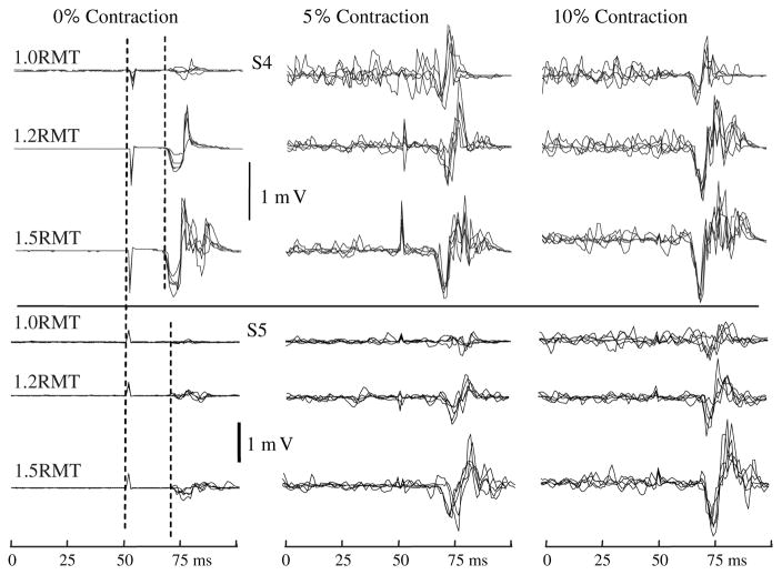
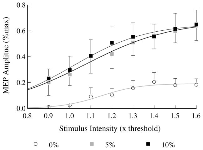
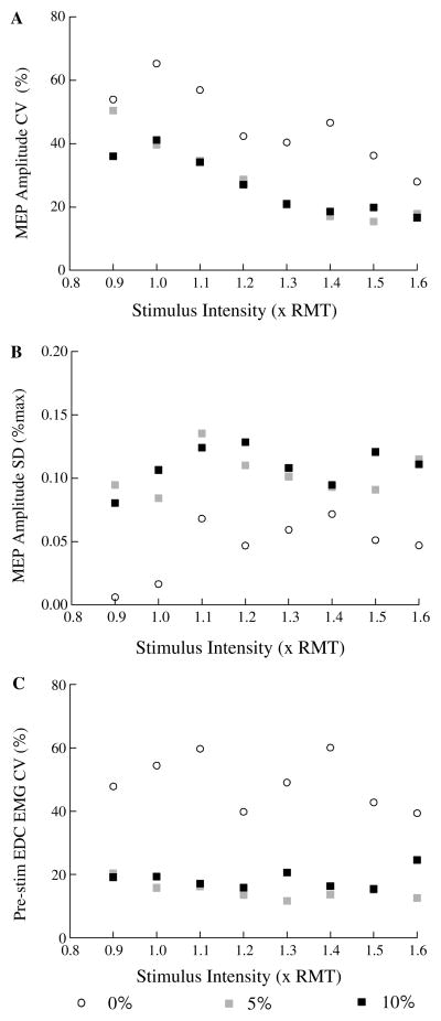
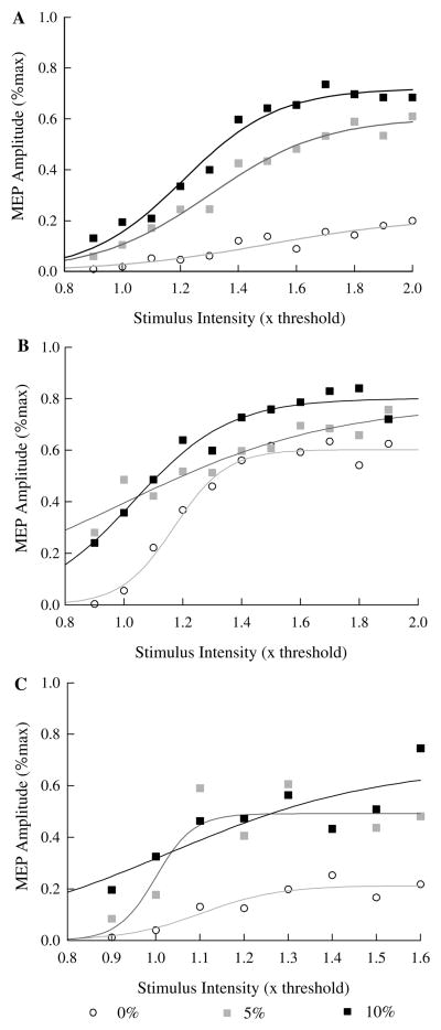
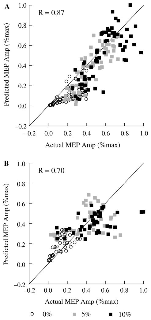
The purpose of this research was to determine whether motor cortex excitability assessed using transcranial magnetic stimulation (TMS) is less variable when subjects maintain a visually controlled low-level contraction of the muscle of interest. We also examined the dependence of single motor evoked potential (MEP) amplitude on stimulation intensity and pre-stimulus muscle activation level using linear and non-linear multiple regression analysis. Eight healthy adult subjects received single pulse TMS over the left motor cortex at a point where minimal stimulation intensity was required to produce MEPs in extensor digitorum communis (EDC). Voluntary activation of the muscle was controlled by visual display of a target force (indicated by a stable line on an oscilloscope) and the isometric force produced as the subject attempted to extend the fingers (indicated by a line on the oscilloscope representing the finger extension force) while subjects were instructed to: exert zero extension force (0%) and produce forces equal to 5 and 10% of maximum voluntary finger extension under separate conditions. Relative variability (coefficient of variation) of single MEPs at a constant stimulus intensity and of pre-stimulus muscle EMG was lower during maintained 5 and 10% contractions than at 0% contraction levels. Therefore, maintaining a stable low intensity contraction helps stabilize cortical and spinal excitability. Multiple regression analyses showed that a linear dependence of single MEPs on stimulation intensity and pre-stimulus muscle activation level produced similar fits to those for a non-linear dependence on stimulus intensity and a linear dependence on pre-stimulus EMG. Thus, a simple linear method can be used to assess dependence of single MEP amplitudes on both stimulus intensity (to characterize slope of the recruitment curve) and low intensity background muscle activation level.
Figures
References
-
- Abbruzzese G, Trompetto C, Schieppati M. The excitability of the human motor cortex increases during execution and mental imagination of sequential but not repetitive finger movements. Exp Brain Res. 1996;111:465–472. - PubMed
-
- Alstermark B, Isa T, Tantisira B. Integration in descending motor pathways controlling the forelimb in the cat. 18. Morphology, axonal projection and termination of collaterals from C3-C4 propriospinal neurones in the segment of origin. Exp Brain Res. 1991a;84:561–568. - PubMed
-
- Alstermark B, Isa T, Tantisira B. Pyramidal excitation in long propriospinal neurones in the cervical segments of the cat. Exp Brain Res. 1991b;84:569–582. - PubMed
-
- Amassian VE, Quirk GJ, Stewart M. A comparison of corticospinal activation by magnetic coil and electrical stimulation of monkey motor cortex.[see comment] Electroencephalogr Clin Neurophysiol. 1990;77:390–401. - PubMed
Publication types
MeSH terms
Grants and funding
LinkOut - more resources
Full Text Sources
