. 2006;20(11):1628-36.
doi: 10.1002/rcm.2482.

Investigating the structural properties of amyloid-like fibrils formed in vitro from beta2-microglobulin using limited proteolysis and electrospray ionisation mass spectrometry

Affiliations

Investigating the structural properties of amyloid-like fibrils formed in vitro from beta2-microglobulin using limited proteolysis and electrospray ionisation mass spectrometry

Sarah L Myers et al. Rapid Commun Mass Spectrom. 2006.

Abstract

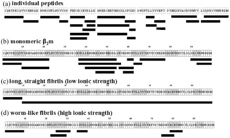
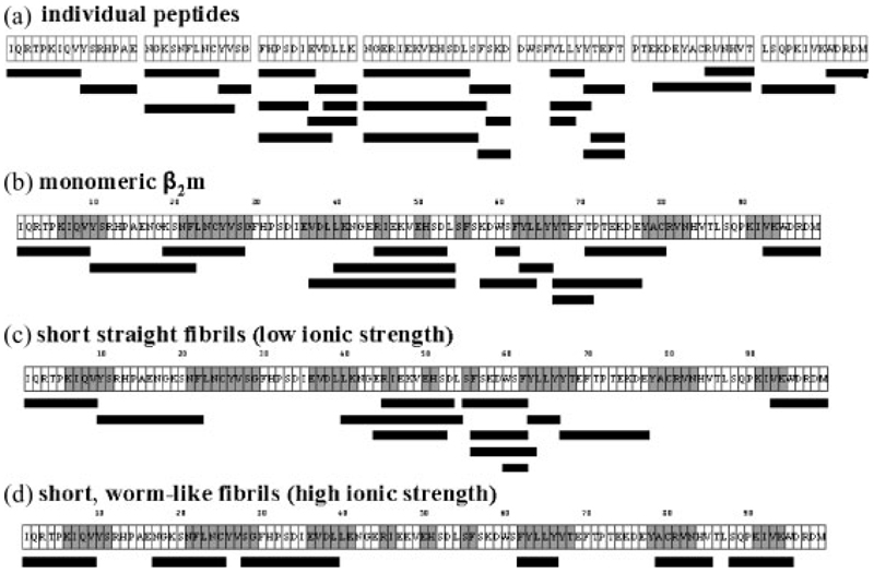
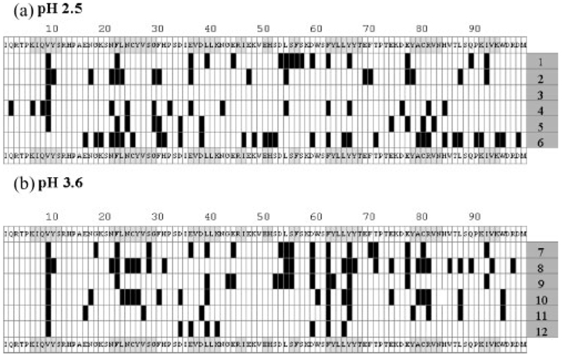
The protein beta(2)-microglobulin (beta(2)m) aggregates to form classical amyloid fibrils in patients undergoing long-term haemodialysis. Amyloid-like fibrils with a cross-beta fold can also be formed from wild-type beta(2)m under acidic conditions in vitro. The morphology of such fibrils depends critically on the conditions used: incubation of beta(2)m in low ionic strength buffers at pH 2.5 results in the formation of long (microm), straight fibrils while, at pH 3.6, short (<500 nm) fibrils form. At higher ionic strengths (0.2-0.4 M) at pH 1.5-3.6, the fibrils have a distinct curved and nodular morphology. To determine the conformational properties of beta(2)m within in vitro fibrils of different morphologies, limited proteolysis of each fibril type using pepsin was performed and the resulting peptide fragments identified by tandem mass spectrometry. For comparison, the proteolytic degradation patterns of monomeric beta(2)m and seven synthetic peptides spanning the entire sequence of the intact protein were similarly analysed. The results show that fibrils with different morphologies result in distinct digestion patterns. While the curved, worm-like fibrils are relatively weakly protected from proteolysis, the long, straight fibrils formed at pH 2.5 at low ionic strength show only a single cut-site at Val9, demonstrating that substantial refolding of the initially acid-denatured and unprotected state of beta(2)m occurs during assembly. The data demonstrate that the organisation of the polypeptide chain in fibrils with different morphological features differs considerably, despite the fact that the fibrils possess a common cross-beta architecture.


Figures

Figure 1
Figure 1
(a) Ribbon diagram of native β2m showing the seven β-strands (labelled A to G) and the disulphide bond between strands B and F (linking Cys25 and Cys80). The figure was drawn using Molscript and Raster3D using the coordinates 1DUZ. (b–f) Tapping-mode AFM images showing: (b) monomeric β2m (pH 7.0); (c–f) fibrils formed by incubating β2m at: (c) pH 2.5 (low ionic strength); (d) pH 2.5 (high ionic strength); (e) pH 3.6 (low ionic strength); and (f) pH 3.6 (high ionic strength).
Figure 2
Figure 2
Limited pepsin proteolysis at pH 2.5 of (a) seven synthetic peptides which together comprise the entire sequence of β2m; (b) monomeric β2m; (c) fibrils formed under low ionic strength conditions; and (d) fibrils formed under high ionic strength conditions. The 99-residue amino acid sequence is shown in (b)–(d) (without the N-terminal initiating Met residue). The regions of grey shading indicate the location of the native β-strands. Black bars under the sequences show the peptide fragments identified by ESI-MS(/MS) after limited proteolysis.
Figure 3
Figure 3
Limited pepsin proteolysis at pH 3.6 of (a) seven synthetic peptides which together comprise the entire sequence of β2m; (b) monomeric β2m; (c) fibrils formed under low ionic strength conditions; and (d) fibrils formed under high ionic strength conditions. The 99-residue amino acid sequence is shown in (b)–(d) (without the N-terminal initiating Met residue). The regions of grey shading indicate the location of β-strands. Black bars under the sequences show the peptide fragments identified by ESI-MS(/MS) after limited proteolysis.
Figure 4
Figure 4
(a) ESI-MS mass spectrum of monomeric β2m after pepsin proteolysis at pH 2.5. The protein was incubated with pepsin (100:1 w/w β2m/pepsin) at 258C for 15 min and the resulting mixture analysed on a Q-Tof mass spectrometer (Waters Corp., Manchester, UK) equipped with a nano-ESI source. The spectrum shows a mixture of singly and multiply charged protonated molecules. The MH+ ion at m/z 571.3 has been circled as an example of a peptide that was subsequently subjected to MS/MS analysis (see (b)). (b) MS/MS analysis of the MH+ ion at m/z 571.3 in (a). MS/MS analyses were performed by fragmentation of the selected precursor ions in the collision cell. The product ions were analysed and the resulting spectra interpreted for sequence information. The a, b, and y″ sequence-specific ions labelled on the spectrum confirm the sequence of Tyr.Leu.Leu.Tyr (residues 63–66; 570.3 Da).
Figure 5
Figure 5. Summary of the cleavage sites observed by ESI-MS(/MS) after pepsin proteolysis.
The rows illustrate data obtained at (a) pH 2.5: (1) and (2) monomeric β2m (15 min and 24 h digestion, respectively), (3) and (4) fibrils formed under low ionic strength conditions (15 min and 24 h digestion, respectively), and (5) and (6) fibrils formed under high ionic strength conditions (15 min and 24 h digestion, respectively); (b) pH 3.6: (7) and (8) monomeric β2m (15 min and 24 h digestion, respectively), (9) and (10) fibrils formed under low ionic strength conditions (15 min and 24 h digestion), and (11) and (12) fibrils formed under high ionic strength conditions (15 min and 24 h digestion, respectively). Limited proteolysis was performed with pepsin/β2m (1:100 w/w) at 258C for 15 min, and extensive proteolysis for 24 h. The 99-residue amino acid sequence is shown (without the N-terminal initiating Met residue). The regions of grey shading indicate the location of β-strands. The vertical bars in (a) and (b) depict the C-terminal residue of an identified peptide fragment and hence indicate the proteolytic cleavage sites.

Similar articles

Cited by

References

    1. Pepys MB. Annu Rev Med. 2006;57:223. - PubMed
    1. Sacchettini JC, Kelly JW. Nature Rev Drug Discov. 2002;1:267. - PubMed
    1. Rochet JC, Landsbury PT. Curr Opin Struct Biol. 2000;10:60. - PubMed
    1. Uversky VN, Fink AL. Biochim Biophys Acta. 2004;1698:131. - PubMed
    1. Fandrich M, Dobson CM. EMBO J. 2002;21:5682. doi: 10.1093/emboj/cdf573. - DOI - PMC - PubMed

Publication types

LinkOut - more resources