

Protocadherin-PC promotes androgen-independent prostate cancer cell growth
- PMID: 16637074
- PMCID: PMC2660890
- DOI: 10.1002/pros.20446
Protocadherin-PC promotes androgen-independent prostate cancer cell growth
Abstract
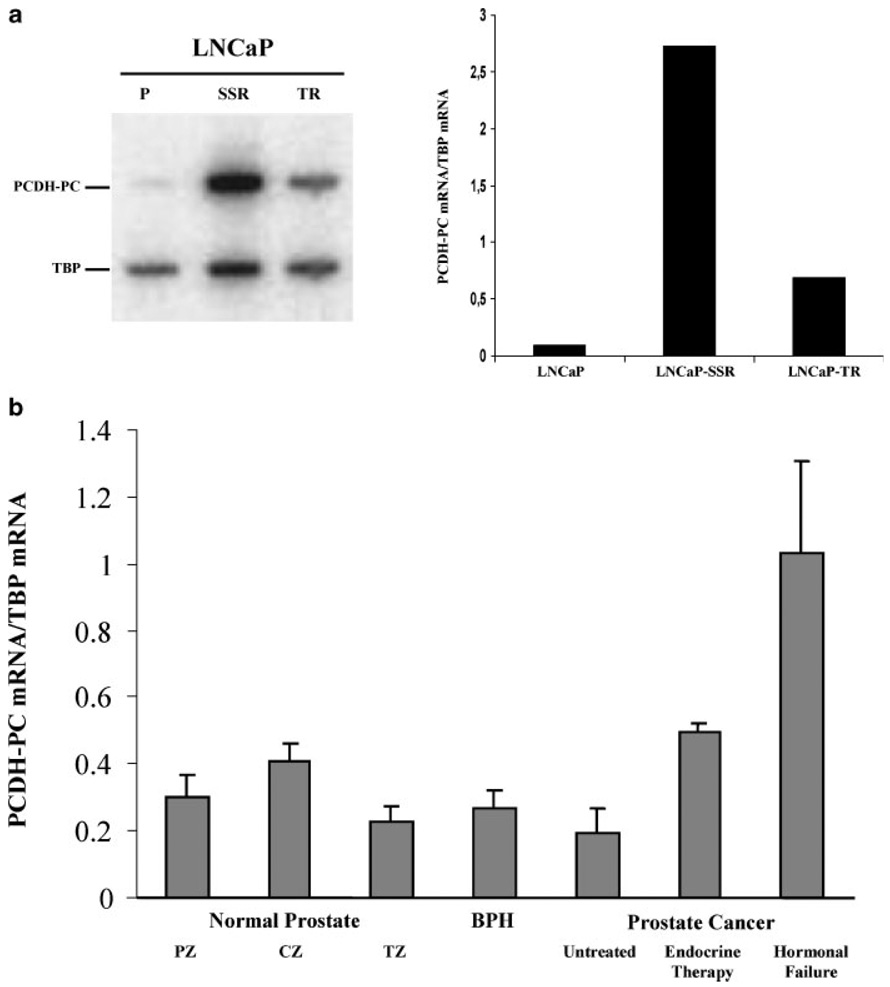
Background: Protocadherin-PC (PCDH-PC) expression is upregulated in apoptosis-resistant sublines of the LNCaP human prostate cancer (CaP) cell line. Here, we assess the role of PCDH-PC in CaP cells and its mRNA expression in human prostate tissues.
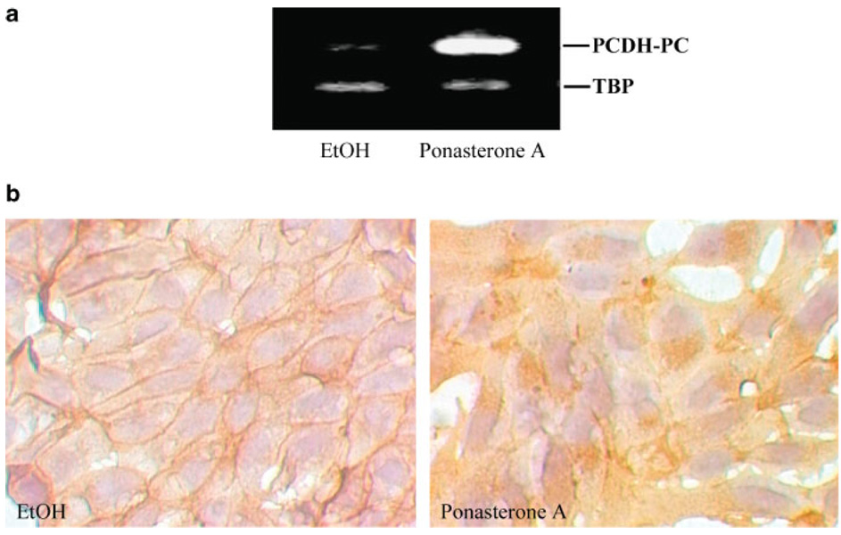
Methods: LNCaP cells transfected with PCDH-PC were tested for their ability to grow in vitro and in vivo in androgen-deprived conditions. PCDH-PC mRNA expression was evaluated by semi-quantitative RT-PCR and by in situ hybridization.
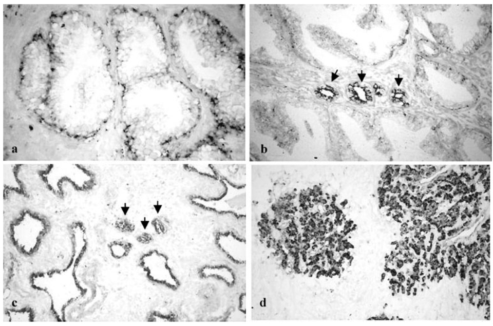
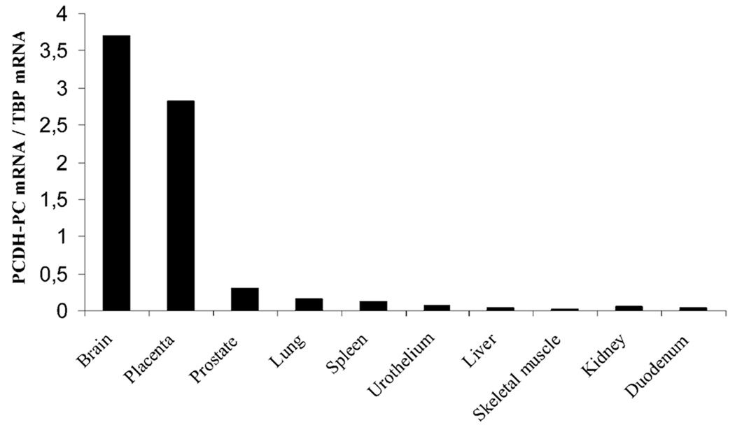
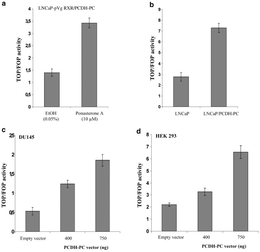
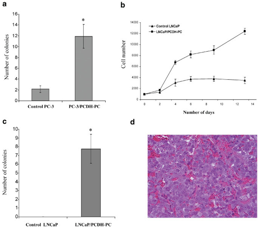
Results: PCDH-PC expression induced Wnt signaling in CaP cells and permitted androgen-independent growth of hormone-sensitive CaP cells. Expression of PCDH-PC-homologous transcripts was low and restricted to some epithelial cells in normal tissue and to CaP cells in tumors. However, hormone-resistant CaP cells expressed significantly higher levels of PCDH-PC-related mRNA.
Conclusions: Our findings suggest a novel mechanism for the progression of CaP involving expression of PCDH-PC. This novel protocadherin induces Wnt signaling, promotes malignant behavior and hormone-resistance of CaP cells.
Copyright 2005 Wiley-Liss, Inc.
Figures
Similar articles
-
A human- and male-specific protocadherin that acts through the wnt signaling pathway to induce neuroendocrine transdifferentiation of prostate cancer cells.Cancer Res. 2005 Jun 15;65(12):5263-71. doi: 10.1158/0008-5472.CAN-05-0162. Cancer Res. 2005. PMID: 15958572
-
The emergence of protocadherin-PC expression during the acquisition of apoptosis-resistance by prostate cancer cells.Oncogene. 2002 Nov 7;21(51):7861-71. doi: 10.1038/sj.onc.1205991. Oncogene. 2002. PMID: 12420223
-
GREB1 is a novel androgen-regulated gene required for prostate cancer growth.Prostate. 2006 Jun 1;66(8):886-94. doi: 10.1002/pros.20403. Prostate. 2006. PMID: 16496412
-
Differential regulation of IGFBP-3 by the androgen receptor in the lineage-related androgen-dependent LNCaP and androgen-independent C4-2 prostate cancer models.Prostate. 2006 Jun 15;66(9):971-86. doi: 10.1002/pros.20420. Prostate. 2006. PMID: 16541420
-
Androgen deprivation induces selective outgrowth of aggressive hormone-refractory prostate cancer clones expressing distinct cellular and molecular properties not present in parental androgen-dependent cancer cells.Cancer J. 2000 Jul-Aug;6(4):220-33. Cancer J. 2000. PMID: 11038142
Cited by
-
DNA methylation alterations at RE1-silencing transcription factor binding sites and their flanking regions in cancer.Clin Epigenetics. 2023 Jun 10;15(1):98. doi: 10.1186/s13148-023-01514-9. Clin Epigenetics. 2023. PMID: 37301955 Free PMC article.
-
Decoding Somatic Driver Gene Mutations and Affected Signaling Pathways in Human Medulloblastoma Subgroups.J Cancer. 2018 Nov 17;9(24):4596-4610. doi: 10.7150/jca.27993. eCollection 2018. J Cancer. 2018. PMID: 30588243 Free PMC article.
-
Non-clustered protocadherin.Cell Adh Migr. 2011 Mar-Apr;5(2):97-105. doi: 10.4161/cam.5.2.14374. Epub 2011 Mar 1. Cell Adh Migr. 2011. PMID: 21173574 Free PMC article. Review.
-
Androgen deprivation leads to increased carbohydrate metabolism and hexokinase 2-mediated survival in Pten/Tp53-deficient prostate cancer.Oncogene. 2017 Jan 26;36(4):525-533. doi: 10.1038/onc.2016.223. Epub 2016 Jul 4. Oncogene. 2017. PMID: 27375016 Free PMC article.
-
Aberrant expression and functions of protocadherins in human malignant tumors.Tumour Biol. 2016 Oct;37(10):12969-12981. doi: 10.1007/s13277-016-5169-9. Epub 2016 Jul 24. Tumour Biol. 2016. PMID: 27449047 Review.
References
-
- Gittes RF. Carcinoma of the prostate. N Engl J Med. 1991;324(4):236–245. - PubMed
-
- Landis SH, Murray T, Bolden S, Wingo PA. Cancer statistics, 1999. CA Cancer J Clin. 1999;49(1):8–31. - PubMed
-
- Schulze H, Isaacs JT, Coffey DS. A critical review of the concept of total androgen ablation in the treatment of prostate cancer. Prog Clin Biol Res. 1987;243A:1–19. - PubMed
-
- Grayhack JT, Keeler TC, Kozlowski JM. Carcinoma of the prostate. Hormonal therapy. Cancer. 1987;60(3 Suppl):589–601. - PubMed
-
- Carter HB, Isaacs JT. Overview of hormonal therapy for prostate cancer. Prog Clin Biol Res. 1990;350:129–140. - PubMed
Publication types
MeSH terms
Substances
Grants and funding
LinkOut - more resources
Full Text Sources
Medical
Miscellaneous
