Review
. 2006 Apr;208(4):471-90.
doi: 10.1111/j.1469-7580.2006.00540.x.

Where tendons and ligaments meet bone: attachment sites ('entheses') in relation to exercise and/or mechanical load

Affiliations
Review

Where tendons and ligaments meet bone: attachment sites ('entheses') in relation to exercise and/or mechanical load

M Benjamin et al. J Anat. 2006 Apr.

Abstract

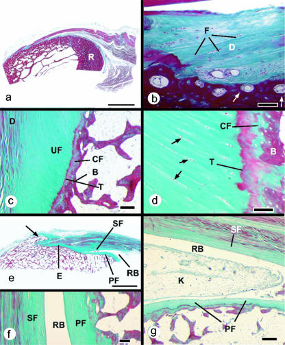
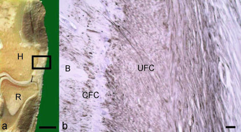
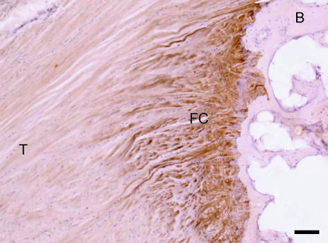
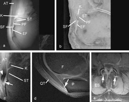
Entheses (insertion sites, osteotendinous junctions, osteoligamentous junctions) are sites of stress concentration at the region where tendons and ligaments attach to bone. Consequently, they are commonly subject to overuse injuries (enthesopathies) that are well documented in a number of sports. In this review, we focus on the structure-function correlations of entheses on both the hard and the soft tissue sides of the junction. Particular attention is paid to mechanical factors that influence form and function and thus to exploring the relationship between entheses and exercise. The molecular parameters indicative of adaptation to mechanical stress are evaluated, and the basis on which entheses are classified is explained. The application of the 'enthesis organ' concept (a collection of tissues adjacent to the enthesis itself, which jointly serve the common function of stress dissipation) to understanding enthesopathies is considered and novel roles of adipose tissue at entheses are reviewed. A distinction is made between different locations of fat at entheses, and possible functions include space-filling and proprioception. The basic anchorage role of entheses is considered in detail and comparisons are explored between entheses and other biological 'anchorage' sites. The ability of entheses for self-repair is emphasized and a range of enthesopathies common in sport are reviewed (e.g. tennis elbow, golfer's elbow, jumper's knee, plantar fasciitis and Achilles insertional tendinopathies). Attention is drawn to the degenerative, rather than inflammatory, nature of most enthesopathies in sport. The biomechanical factors contributing to the development of enthesopathies are reviewed and the importance of considering the muscle-tendon-bone unit as a whole is recognized. Bony spur formation is assessed in relation to other changes at entheses which parallel those in osteoarthritic synovial joints.


Figures

Fig. 1
Fig. 1
Histological sections (stained with Masson's trichrome) of entheses from dissecting room cadavers. (a) A macroscopic view of the fibrous enthesis at the insertion of pronator teres on the mid-shaft of the radius (R). Note the thick layer of cortical bone. Scale bar = 5 mm. (b) Higher magnification view of the pronator teres enthesis, showing the presence of dense fibrous connective tissue (D) at the bone–tendon junction. Fibroblasts are evident (F), but no fibrocartilage cells. Note the presence of osteons at the attachment site (arrows). Scale bar = 100 µm. (c) A typical fibrocartilaginous enthesis (Achilles tendon) showing the four zones of tissue at the bone–tendon interface: dense fibrous connective tissue (D), uncalcified fibrocartilage (UF), calcified fibrocartilage (CF) and bone (B). The two fibrocartilage zones are separated from each other by a tidemark (T). Note that the tidemark is straight, but the tendon–bone interface (i.e. the junction between calcified and non-calcified fibrocartilage) is highly irregular (arrows). This is critical for the integrity of the junction. Scale bar = 300 µm. (d) A higher magnification view of the fibrocartilage zones at the Achilles tendon enthesis. The zone of uncalcified fibrocartilage is characterized by rows of fibrocartilage cells (arrows) that are separated from each other by parallel bundles of collagen fibres. T, tidemark; B, bone. Scale bar = 100 µm. (e) A macroscopic view of the Achilles tendon enthesis organ. This consists of the enthesis itself (E), a thick periosteal fibrocartilage (PF) on the superior tuberosity (ST) of the calcaneus, a sesamoid fibrocartilage (SF) in the deep surface of the tendon, an intervening retrocalcaneal bursa (RB) and the tip of Kager's fat pad (not visible in this section). Note the enthesophyte (arrow) in the most inferior part of the enthesis. Scale bar = 5 mm. (f) A higher magnification view of the sesamoid (SF) and periosteal (PF) fibrocartilages that lie either side of the retrocalcaneal bursa (RB). Scale bar = 100 µm. (g) The wedge-shaped tip of Kager's fat pad (K) protrudes into the retrocalcaneal bursa (RB) in a plantarflexed foot. PF, periosteal fibrocartilage; SF, sesamoid fibrocartilage. Scale bar = 500 µm.
Fig. 2
Fig. 2
(a) Frontal saw cut through the lateral part of a human elbow joint. The rectangle represents the part of the common extensor origin that is shown in fig. b. H, humerus; R, radius. Scale bar = 1 cm. (b) Immunohistochemical labelling for aggrecan at the enthesis of the common extensor origin. B, bone; UFC, uncalcified fibrocartilage; CFC, calcified fibrocartilage. Scale bar = 100 µm.
Fig. 3
Fig. 3
Immunohistochemical labelling for type II collagen at the insertion of the central slip of the extensor tendon to the base of the middle phalanx of a second toe. The enthesis fibrocartilage (FC) has labelled strongly for type II collagen, but the bone (B) and tendon (T) are not labelled. Scale bar = 100 µm.
Fig. 4
Fig. 4. MR images of entheses
(a) A UTE image and (b) a corresponding methacrylate section of the Achilles tendon (AT) enthesis, cut in the mid-sagittal plane to show the periosteal (PF), sesamoid (SF) and enthesis (EF) fibrocartilages, which can be identified in the UTE sequence. K, Kager's fat pad; ST, superior tuberosity. Panel b is reproduced with permission from Robson et al. Clin Radiol 2004, 59, 729. (c) A sagittal magic angle image of the Achilles tendon enthesis. Low signal is seen in the tendon posterior to the calcaneus (arrowhead), but high signal is seen in the sesamoid fibrocartilage (arrow). (d) Sagittal magic angle image of the quadriceps tendon (QT) insertion on the patella (P). The enthesis fibrocartilage (arrow) has a high signal. F, femur. (e) Tranverse UTE image of the lumbar spine in a patient with degenerative spine disease. High signal is seen in the region of the dorsal capsule of the lumbar facet joints (arrows). ES, erector spinae.

Similar articles

Cited by

References

    1. Abreu MR, Chung CB, Mendes L, Mohana-Borges A, Trudell D, Resnick D. Plantar calcaneal enthesophytes: new observations regarding sites of origin based on radiographic, MR imaging, anatomic, and paleopathologic analysis. Skeletal Radiol. 2003;32:13–21. - PubMed
    1. Adams CMW, Bayliss OB, Baker RWR, Abdulla YH, Huntercraig CJ. Lipid deposits in aging human arteries, tendons, and fascia. Atherosclerosis. 1974;19:429–440. - PubMed
    1. Andriacchi TP, Birac D. Functional testing in the anterior cruciate ligament-deficient knee. Clin Orthop Relat Res. 1993;288:40–47. - PubMed
    1. Aoki M, Oguma H, Fukushima S, Ishii S, Ohtani S, Murakami G. Fibrous connection to bone after immediate repair of the canine infraspinatus: the most effective bony surface for tendon attachment. J Shoulder Elbow Surg. 2001;10:123–128. - PubMed
    1. Bell E, Gosline J. Mechanical design of mussel byssus: material yield enhances attachment strength. J Exp Biol. 1996;199:1005–1017. - PubMed

MeSH terms