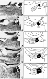
Total number of neurons in the habenular nuclei of the rat epithalamus: a stereological study
- PMID: 16637880
- PMCID: PMC2100216
- DOI: 10.1111/j.1469-7580.2006.00573.x
Total number of neurons in the habenular nuclei of the rat epithalamus: a stereological study
Abstract
The total number of neurons in the medial and lateral habenular nuclei of the rat epithalamus was estimated using modern stereological counting methods and systematic random sampling techniques. Six to eight young adult male rats, and a complete set of serial 40-microm glycolmethacrylate sections for each rat, were used to quantify neuronal numbers. After a random start, a systematic subset (e.g. every third) of the serial sections was used to estimate the total volume of each nucleus using Cavalieri's method. The same set of sampled sections was used to estimate the number of neurons in a known subvolume (i.e. the numerical density N(v)) by the optical disector method. Multiplication of the total volume by N(v) yielded the total number of neurons. It was found that the right medial habenular nucleus consisted, on average, of 18,000 neurons (with a coefficient of variation of 0.18), while the right lateral habenular nucleus had 13,000 neurons on average (0.14). These total neuronal numbers provide important data for the transfer of information through these nuclei and for species comparisons.
Figures


References
-
- Aghajanian GK, Wang RY. Habenular and other midbrain raphe afferents demonstrated by a modified retrograde tracing technique. Brain Res. 1977;122:229–242. - PubMed
-
- Ashwell KWS, Zhang LL. Forebrain hypoplasia following acute prenatal ethanol exposure: quantitative analysis of effects on specific forebrain nuclei. Pathology. 1996;28:161–166. - PubMed
-
- Bjugn R, Gundersen HJG. Estimate of the total number of neurons and glial and endothelial cells in the rat spinal cord by means of the optical disector. J Comp Neurol. 1993;328:406–414. - PubMed
-
- Brændgaard H, Evans SM, Howard CV, Gundersen HJG. Total number of neurons in the human neocortex unbiasedly estimated using optical disectors. J Microsc. 1990;157:285–304. - PubMed
-
- Chadi G, Møller A, Rosén L, et al. Protective actions of human recombinant basic fibroblast growth factor on MPTP-lesioned nigrostriatal dopamine neurons after intraventricular infusion. Exp Brain Res. 1993;97:145–158. - PubMed
MeSH terms
LinkOut - more resources
Full Text Sources

