. 2006 Jun;97(5):1301-13.
doi: 10.1111/j.1471-4159.2006.03797.x. Epub 2006 Apr 21.

Cyclopentenone isoprostanes are novel bioactive products of lipid oxidation which enhance neurodegeneration

Affiliations

Cyclopentenone isoprostanes are novel bioactive products of lipid oxidation which enhance neurodegeneration

Erik S Musiek et al. J Neurochem. 2006 Jun.

Abstract

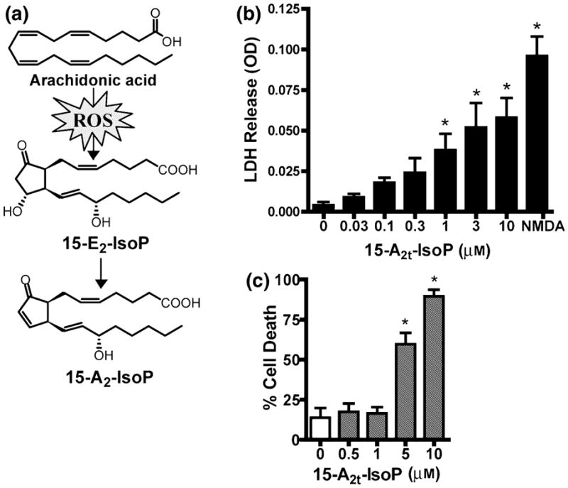
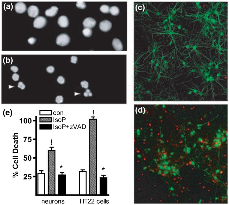
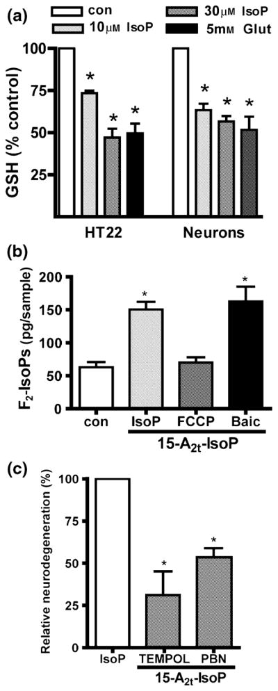
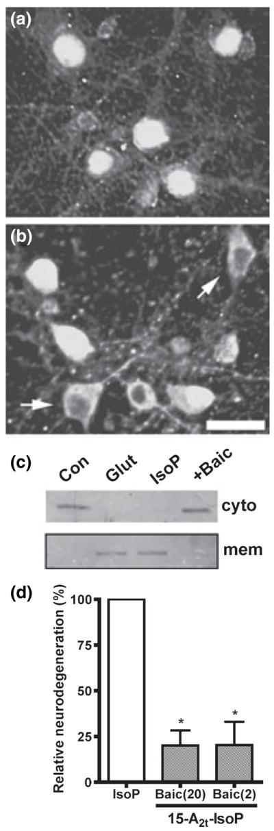
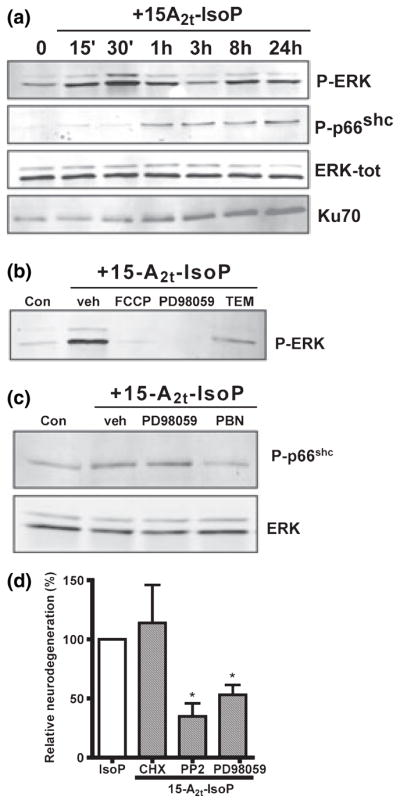
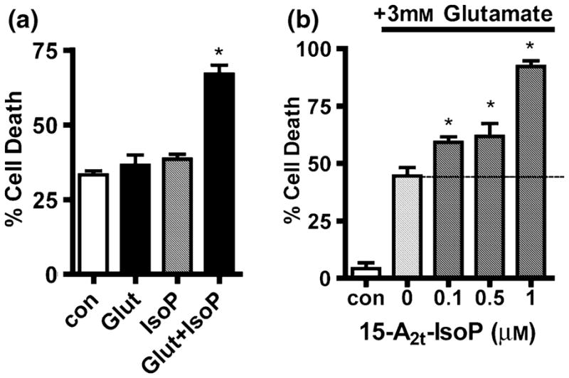
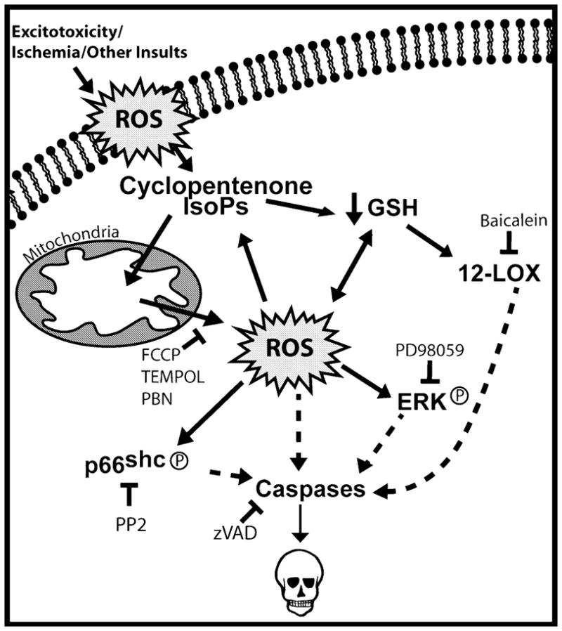
Oxidative stress and subsequent lipid peroxidation are involved in the pathogenesis of numerous neurodegenerative conditions, including stroke. Cyclopentenone isoprostanes (IsoPs) are novel electrophilic lipid peroxidation products formed under conditions of oxidative stress via the isoprostane pathway. These cyclopentenone IsoPs are isomeric to highly bioactive cyclopentenone prostaglandins, yet it has not been determined if these products are biologically active or are formed in the brain. Here we demonstrate that the major cyclopentenone IsoP isomer 15-A2t-IsoP potently induces apoptosis in neuronal cultures at submicromolar concentrations. We present a model in which 15-A2t-IsoP induced neuronal apoptosis involves initial depletion of glutathione and enhanced production of reactive oxygen species, followed by 12-lipoxygenase activation and phosphorylation of extracellular signal-regulated kinase 1/2 and the redox sensitive adaptor protein p66shc, which results in caspase-3 cleavage. 15-A2t-IsoP application also dramatically potentiates oxidative glutamate toxicity at concentrations as low as 100 nm, demonstrating the functional importance of these molecules in neurodegeneration. Finally, we employ novel mass spectrometric methods to show that cyclopentenone IsoPs are formed abundantly in brain tissue under conditions of oxidative stress. Together these findings suggest that cyclopentenone IsoPs may contribute to neuronal death caused by oxidative insults, and that their activity should perhaps be addressed when designing neuroprotective therapies.


Figures

Fig. 1
Fig. 1
15-A2t-Isoprostane (15-A2t-IsoP) is toxic to neurons. (a) Diagram depicting the formation of 15-A2t-IsoP via the oxidation of arachidonic acid and subsequent spontaneous dehydration of 15-E2t-IsoP. (b) Mature primary cortical neurons were exposed to increasing concentrations of 15-A2t-IsoP for 48 h, at which time cell death was assessed by measuring release of lactate dehydrogenase (LDH) into the cell media from dying neurons. NMDA (100 μM) was used as a positive control, as it induces total cell death in these cultures. Note that 15-A2t-IsoP induces cell death in a dose-dependent manner with an LD50 of 950 nM. (c) The oxidative stress-sensitive HT22 hippocampal cell line was exposed to 15-A2t-IsoP for 18 h, after which cell viability was assessed by measuring cellular MTT reducing capacity. Like primary neurons, HT22 cells are killed by 15-A2t-IsoP with an LD50 of 3 μM. Data in (b) and (c) represent the mean ± SEM of at least three separate experiments, each performed at least in triplicate. *p < 0.05 vs. control by one-way ANOVA.
Fig. 2
Fig. 2
15-A2t-Isoprostane (15-A2t-IsoP) induces apoptotic neuronal death. (a, b) 4′-6-Diamidino-2-phenylindole (DAPI) staining of neurons treated with vehicle (a) or 30 μM 15-A2t-IsoP for 24 h (b) demonstrates an increase in asymmetric chromatin formations (arrows), an indication of apoptosis. (c, d) Neuronal culture treated for 48 h with vehicle (c) vs. 30 μM 15-A2t-IsoP (d). IsoP-treated cells (d) show a loss of microtubule-associated protein 2 (MAP-2) staining (green) and increased activated caspase-3 expression (red). Images in (a)–(d) are representative of three separate experiments. (e) Co-incubation of cells [primary cortical neurons (left) or HT22 cells (right)] with 20 μM zVAD-FMK, a broad-spectrum caspase inhibitor, caused a significant reduction in cell death following 24 h treatment with 15-A2t-IsoP as assessed by lactate dehydrogenase (LDH) release (neurons) or MTT assay (HT22s). Data represent the mean + SEM of four independent experiments, each performed in triplicate and analyzed by two-tailed paired t-test. !p < 0.05 vs. control, *p < 0.05 vs. IsoP + zVAD.
Fig. 3
Fig. 3
Glutathione depletion and oxidative stress contribute to 15-A2t-isoprostane (15-A2t-IsoP) toxicity. (a) Glutathione (GSH) levels were measured spectrophotometrically after immature neurons or HT22 cells were exposed for 3 h to vehicle (0.1% dimethylsulfoxide), 10 or 30 μM 15-A2t-IsoP, or 5 mM glutamate, which was used as a positive control. Data represent the mean + SEM of three separate experiments and was analyzed by two-tailed paired t-test. *p < 0.05 vs. control. (b) Neuronal cultures were exposed to vehicle or 15-A2t-IsoP (30 μM) + the mitochondrial uncoupling agent carbonyl cyanide p-trifluoromethoxyphenylhydrazone (FCCP: 10 μM) or baicalein (2 μM) for 3 h, then harvested and assayed for total F2-isoprostane content (F2-IsoPs) by gas chromatography/mass spectrometry (GC/MS). FCCP and baicalein were present 30 min before and during 15-A2t-IsoP exposure. Data represent the mean + SEM for three independent experiments, and were analyzed one-way ANOVA. *p < 0.05 vs. control. (c) Neurons were exposed to 15-A2t-IsoP (30 μM) + the antioxidants TEMPOL (1 mM) or N-tert-butyl-α-phenylnitrone (PBN, 0.5 mM). After 20–24 h, cell death was assessed by lactate dehydrogenase assay. Antioxidants were present for 30 min before and during IsoP exposure, and significantly decreased 15-A2t-IsoP-induced cell death. Data were normalized to the amount of cell death caused by 15-A2t-IsoP alone minus that caused by vehicle (100% relative neurodegeneration) in this and subsequent figures. Data represent the mean + SEM for three separate experiments, each performed in quadruplicate, and were analyzed by one-way ANOVA.*p < 0.05 vs. control (a, b) or IsoP alone (c).
Fig. 4
Fig. 4
12-Lipoxygenase (12-LOX) activation in neurons following 15-A2t-isoprostane (15-A2t-IsoP) exposure causes cell death. Neurons were exposed to (a) vehicle or (b) 30 μM 15-A2t-IsoP for 3 h then fixed, stained with an anti-12-LOX antibody, and subjected to fluorescent microscopy. Translocation of 12-LOX to the membrane is associated with its activation (arrowheads). Photomicrographs are representative of results from three independent experiments. (c) Immature neurons were exposed to vehicle (Con), 30 μM 15-A2t-IsoP + baicalein (Baic: 20 μM) or 5 mM glutamate (Glut, used as a positive control) for 3 h, then membrane and cytoplasmic fractions were prepared and analyzed for 12-LOX by western blot. Blots are representative of two independent experiments. (d) Neurons were exposed to 15-A2t-IsoP (30 μM) + the specific 12-LOX inhibitor baicalein (Baic: 20 or 2 μM). After 20–24 h, cell death was assessed by lactate dehydrogenase (LDH) assay. Baicalein was present for 30 min before and during IsoP exposure. Data represents the mean + SEM for at least three independent experiments, and was analyzed by one-way ANOVA. *p < 0.05 vs. IsoP alone.
Fig. 5
Fig. 5
15-A2t-Isoprostane (15-A2t-IsoP) neurotoxicity involves phosphorylation of extracellular signal-regulated kinase (ERK) and p66shc and up-regulation of Ku70. (a) Neuronal cultures were exposed to 30 μM 15-A2t-IsoP or vehicle for various amounts of time, then whole- cell extracts were harvested and subjected to western blot analysis. ERK is displayed as a loading control. (b) Neurons were exposed to 30 μM 15-A2t-IsoP + the MEK inhibitor PD98059 (20 μM), TEMPOL (1 mM), or carbonyl cyanide p-trifluoromethoxyphenylhydrazone (FCCP: 1 μM), then harvested at 20 h and subjected to western blot using a phospho-ERK specific antibody. (c) Neurons were exposed to 30 μM 15-A2t-IsoP + PD98059 (20 μM), N-tert-butyl-α-phenylnitrone (PBN: 0.5 mM), or baicalein (20 μM), then harvested at 1 h and subjected to western blot analysis. (d) Neurons were exposed to 15-A2t-IsoP (30 μM) + cyclohexamide (3.5 μM), the Src kinase inhibitor PP2 (50 nM), or the MEK inhibitor PD98059 (20 μM). Cell death was assessed after 20–24 h by lactate dehydrogenase (LDH) assay. Data represent the mean ± SEM of three independent experiments; each performed in triplicate, and were analyzed by one-way ANOVA. Blots shown in (a), (b) and (c) are representative of at least three independent experiments. Inhibitors in (b), (c) and (d) were present for 30 min before and during IsoP exposure. PD98059, 2′-amino-3′-methoxyflavone; PP2, 4-amino-5-(4-chlorophenyl)-7-(t-butyl)pyrazolo-[3,4-d]pyrimidine.
Fig. 6
Fig. 6
15-A2t-Isoprostane (15-A2t-IsoP) potentiates oxidative glutamate toxicity in neurons and HT22 cells. (a) Immature (1–2 DIV) primary cortical neurons were exposed to 1.5 mM glutamate or 5 μM 15-A2t-IsoP alone or in combination. Cell death was assessed after 18 h by lactate dehydrogenase (LDH) assay. (b) HT22 cells were exposed to glutamate plus increasing concentrations of 15-A2t-IsoP. Cell viability was assessed by MTT assay after 20–24 h. Note that in both (a) and (b), 15-A2t-IsoP and glutamate in combination resulted in significantly increased cell death. Data in (a) and (b) represent the mean ± SEM of three independent experiments, each performed in triplicate, and were analyzed by one-way ANOVA. *p < 0.05 vs. control.
Fig. 7
Fig. 7
Model of signaling events in 15-A2t-isoprostane (15-A2t-IsoP)- induced neurodegeneration. Based on our findings in this work, we suggest that 15-A2t-IsoPs are formed in membranes (plasma membrane is depicted, but other membranes are included) following an initial oxidant injury. 15-A2t-IsoPs could then induce mitochondrial dysfunction and glutathione (GSH) depletion, causing marked increases in intracellular reactive oxygen species (ROS) production and further lipid peroxidation. This results in phosphorylation of extracellular signal-regulated kinase 1/2 (ERK1/2) and serine 36 of p66shc, both of which contribute to cell death. The loss of GSH leads to the translocation and activation of 12-lipoxygenase (12-LOX), which contributes to cell death. As 15-A2t-IsoP is a product and inducer of oxidative stress, the production of 15-A2t-IsoPs in response to an initial oxidant injury sets in motion a feed-forward cycle that leads to a loss of intracellular redox homeostasis and cell death. Interruption of this cycle with baicalein, 4-amino-5-(4-chlorophenyl)-7-(t-butyl) pyrazolo[3,4-d]pyrimidine (PP2), or antioxidants can prevent cell death. At the earliest stage, inhibition of mitochondrial function with carbonyl cyanide p-trifluoromethoxyphenylhydrazone (FCCP) blocks 15-A2t-IsoP-induced oxidative injury and ERK phosphorylation.

References

    1. Alessandrini A, Namura S, Moskowitz MA, Bonventre JV. MEK1 protein kinase inhibition protects against damage resulting from focal cerebral ischemia. Proc Natl Acad Sci USA. 1999;96:12 866–12 869. - PMC - PubMed
    1. Arai K, Nishiyama N, Matsuki N, Ikegaya Y. Neuro-protective effects of lipoxygenase inhibitors against ischemic injury in rat hippocampal slice cultures. Brain Res. 2001;904:167–172. - PubMed
    1. Balamurugan K, Rajaram R, Ramasami T, Narayanan S. Chromium (III) – induced apoptosis of lymphocytes: death decision by ROS and Src-family tyrosine kinases. Free Radic Biol Med. 2002;33:1622–1640. - PubMed
    1. Beal MF. Aging, energy, and oxidative stress in neurodegenerative diseases. Ann Neurol. 1995;38:357–366. - PubMed
    1. Bruckner SR, Perry G, Estus S. 4-hydroxynonenal contributes to NGF withdrawal-induced neuronal apoptosis. J Neurochem. 2003;85:999–1005. - PubMed

Publication types

MeSH terms