Review
. 2006 May;47(5):1736-45.
doi: 10.1167/iovs.05-1291.

Eye cancer: unique insights into oncogenesis: the Cogan Lecture

Affiliations
Review

Eye cancer: unique insights into oncogenesis: the Cogan Lecture

J William Harbour. Invest Ophthalmol Vis Sci. 2006 May.
No abstract available


Figures

Figure 1
Figure 1
The structure of the Rb pocket domain. Cartoon based on the crystal structure of the highly conserved A and B boxes of Rb, which form a strong intramolecular complex. The tubular structures represent peptide segments from E2F, which binds Rb at a cleft formed between the A and B boxes, and the human papillomavirus E7 protein, which binds Rb at a site in the B box through its LxCxE motif. Adapted, with permission, from Lee C, Chang JH, Lee HS, Cho Y. Structural basis for the recognition of the E2F transactivation domain by the retinoblastoma tumor suppressor. Genes Dev. 2002;16:3199–3212. © 2002 Cold Spring Harbor Laboratory Press.
Figure 2
Figure 2
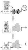
Mechanisms of transcriptional repression by Rb. Rb represses transcription by recruiting chromatin remodeling proteins such as histone deacetylases (HDAC) and BRG1 to DNA regulatory elements containing E2F sites through its interaction with E2F proteins. The resulting alterations in chromatin structure prevent access by the transcriptional machinery. Phosphorylation of Rb by cyclin dependent kinases removes these chromatin remodeling proteins from the multimeric complex, thereby allowing expression of genes involved in cell division. More complete phosphorylation or mutational loss of Rb releases the transactivation domain of E2F to directly activate genes, especially those involved in apoptosis.
Figure 3
Figure 3
Progressive phosphorylation of Rb. Rb is progressively phosphorylated by successive cyclin-CDK complexes, which allows the consecutive derepression of cyclins during cell cycle progression. Thus, Rb may serve to regulate the orderly cell cycle progression, which could explain why Rb-null cells are prone to chromosomal instability.
Figure 4
Figure 4
The Rb cell fate checkpoint. Rb regulates a major checkpoint in the cell fate decision between proliferation, differentiation, and apoptosis. Partial phosphorylation of Rb allows cell division without triggering an apoptotic response. Hypophosphorylation of Rb allows it to interact with chromatin remodeling proteins such as HDAC and BRG1, which induces widespread repression of specific genes that leads to cell cycle exit, such as occurs in differentiation and senescence. Ultimate phosphorylation of Rb at Ser567, which may occur only during abnormal hyperproliferation or stress conditions, disrupts the Rb protein, leading to its degradation. This also releases free E2F1 to induce the expression of proapoptotic genes.
Figure 5
Figure 5
The role of Rb in melanocyte differentiation. (A) The melanocyte differentiation factor MITF transcriptionally activates the p16Ink4a tumor suppressor, which activates Rb by inhibiting CDK4. Active Rb then enforces cell cycle exit at the G1-S boundary, which is required for efficient melanocyte differentiation. (B) Melanocyte differentiation in vivo is hindered when p16Ink4a is not present to activate Rb. Low-power photomicrographs show immunofluorescence analysis of the melanocyte markers S100, DCT, and TRP1 in cutaneous hair follicles of 3-week-old INK4A-wild-type mice and INK4A-null mice. Note that the expression of S100 (green), TRP1 (red), and DCT (red) is greatly diminished in INK4A-null mice. DAPI (blue) indicates cell nuclei. Scale bar, 100 μm.
Figure 6
Figure 6
Two molecular classes of uveal melanoma. (A) Unsupervised analysis of primary uveal melanomas using principal component analysis reveals two distinct groups of uveal melanomas based on gene expression patterns. (B) Hierarchical clustering of the top 62 genes that discriminate between class 1 and 2 melanomas shows that the two tumor classes have distinct “molecular signatures.” (C) Microarray-based comparative genomic hybridization experiments show that the class 2 signature is strongly associated with monosomy 3 (down arrows) and inversely associated with gain of chromosome 6p (up arrows). Reprinted, with permission, from Onken MD, Worley LA, Ehlers JP, Harbour JW. Gene expression profiling in uveal melanoma reveals two molecular classes and predicts metastatic death. Cancer Res. 2004;64:7205–7209. © 2004 American Association for Cancer Research.
Figure 7
Figure 7
Provisional model of the major genetic events in uveal melanoma. Re-entry of uveal melanocytes into the cell cycle by disruption of the Rb pathway is probably an early event. Limited proliferation may then lead to a population of low-grade neoplastic melanocytes that clinically would be recognized as a nevus. Further mutations that inhibit apoptosis, induce angiogenesis, alter cell-cell and cell–matrix adhesion, alter immunogenicity and dispense with constraints of the differentiated phenotype are probably necessary for the tumor to progress further toward malignancy. Finally, there is a major bifurcation as the melanoma exhibits either a class 1 or 2 molecular profile. Currently, it is not known whether class 1 tumors evolve into class 2 tumors, or whether they progressed along different lines from a very early point in tumor development. Cartoon of a normal eye adapted from image #NEA05 from the Web site of the National Eye Institute, National Institutes of Health.

Similar articles

Cited by

References

    1. Harbour JW. Retinoblastoma: pathogenesis and diagnosis. In: Char DH, editor. Tumors of the Eye and Orbit. Philadelphia: BC Decker; 2001. pp. 253–265.
    1. Neel JV, Falls HF. The rate of mutation of the gene responsible for retinoblastoma in man. Science. 1951;114:419–422. - PubMed
    1. Knudson AG., Jr Mutation and cancer: statistical study of retinoblastoma. Proc Natl Acad Sci USA. 1971;68:820–823. - PMC - PubMed
    1. Friend SH, Bernards R, Rogelj S, et al. A human DNA segment with properties of the gene that predisposes to retinoblastoma and osteosarcoma. Nature. 1986;323:643–646. - PubMed
    1. Lee C, Chang JH, Lee HS, Cho Y. Structural basis for the recognition of the E2F transactivation domain by the retinoblastoma tumor suppressor. Genes Dev. 2002;16:3199–3212. - PMC - PubMed

Publication types

MeSH terms

Substances