. 2006 May 2;103(18):7024-9.
doi: 10.1073/pnas.0602266103. Epub 2006 Apr 25.

Pre-B cell proliferation and lymphoblastic leukemia/high-grade lymphoma in E(mu)-miR155 transgenic mice

Affiliations

Pre-B cell proliferation and lymphoblastic leukemia/high-grade lymphoma in E(mu)-miR155 transgenic mice

Stefan Costinean et al. Proc Natl Acad Sci U S A. .

Abstract

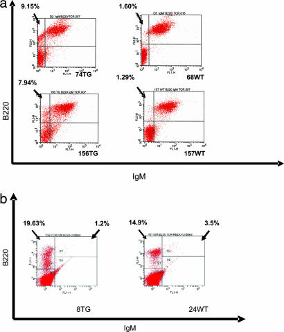
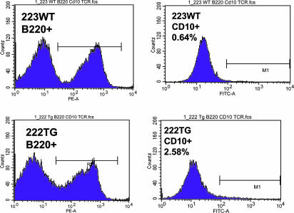
MicroRNAs (miRNAs) represent a newly discovered class of posttranscriptional regulatory noncoding small RNAs that bind to targeted mRNAs and either block their translation or initiate their degradation. miRNA profiling of hematopoietic lineages in humans and mice showed that some miRNAs are differentially expressed during hematopoietic development, suggesting a role in hematopoietic cell differentiation. In addition, recent studies suggest the involvement of miRNAs in the initiation and progression of cancer. miR155 and BIC, its host gene, have been reported to accumulate in human B cell lymphomas, especially in diffuse large B cell lymphomas, Hodgkin lymphomas, and certain types of Burkitt lymphomas. Here, we show that E(mu)-mmu-miR155 transgenic mice exhibit initially a preleukemic pre-B cell proliferation evident in spleen and bone marrow, followed by frank B cell malignancy. These findings indicate that the role of miR155 is to induce polyclonal expansion, favoring the capture of secondary genetic changes for full transformation.


Conflict of interest statement

Conflict of interest statement: No conflicts declared.

Figures

Fig. 1.
Fig. 1.
Production and characterization of Eμ-mmu-miR155. (a) A construct for the miR155 transgene was designed as shown. The mmu-miR155 was cloned between the EcoRV and SalI sites, putting the transgene under the control of the VH promoter Eμ enhancer. The construct was then injected in the male pronuclei of the oocytes of pregnant C57BL/6 and FVB/N female mice. (b) Southern blot was used to genotype the founders. Fifteen transgenic founders were born, seven on a C57BL/6 background (b Left, lanes 1, 3, 5, 6, 7, 10, and 14 are transgenics, and lanes 2, 4, 8, 9, 11, 12, 13, and 15 are wild types) and eight on an FVB/N background (b Right, lanes 1, 3, 5, 7, 9, 11, 13, and 15 are transgenics, and lanes 2, 4, 6, 8, 10, 12, and 14 are wild types). These were then bred to wild-type strain-matched mice to produce 15 transgenic lines. (c) In each of the 15 transgenic lines, expression of the transgene was assessed by Northern blot on total RNA extracted from the lymphocytes isolated from the spleens of 3-week-old mice by using the antisense oligonucleotide of the mmu-miR155 mature sequence as a probe. Five of the transgenic lines with the highest level of expression of the mature miR155 in the splenocytes were selected for further breeding and analysis; one transgenic line did not express the transgene [lanes 1, 2, 3, 5, 8, and 9 (transgenics); lanes 4, 6, and 7 (wild types)].
Fig. 2.
Fig. 2.
Transgenic mice, 6 months old, presented an enlarged abdomen and important splenomegaly. (A) Transgenic mice, 6 months old, had a considerably enlarged abdomen compared with wild-type mice, due to the clinically evident splenomegaly. (B) Spleens of the mice shown in A. The transgenic spleen is enlarged due to expansion of leukemic/lymphoma cells.
Fig. 3.
Fig. 3.
Histology and immunohistochemistry of transgenic spleens compared with the wild type. (a) H&E, spleen (×200), atypical lymphoid proliferation compressing the white pulp in mouse no. 50 (founder 10), 3 weeks old. (b) H&E, spleen (×100), founder no. 8, 6 months of age. The overall architecture of the spleen is being replaced by the atypical lymphoid proliferation. There are only a few remaining germinal lymphoid follicles, greatly decreased in size, compressed by the proliferation. (c) H&E, spleen (×200), transgenic mouse, founder line no. 8, 6 months old. The spleen architecture has been almost completely effaced by the lymphoblastic proliferation. There are still visible remnants of two small compressed lymphoid follicules. (d) H&E, bone marrow (×400), transgenic mouse, founder line no. 8, 6 months old showing the lymphoblastic proliferation in the bone marrow that leads to the replacement of the hematopoietic foci. (e) H&E, normal spleen (×200). (f) Immunohistochemistry, spleen, transgenic mouse no. 72, 3 weeks old, for Ki67, showing increased lymphoid proliferation in the spleen (×200).
Fig. 4.
Fig. 4.
IgM staining of the atypical lymphoid proliferation in the spleen of a transgenic mouse, 3 weeks old. IgM is present in the cytoplasm of the proliferating lymphocytes (cIgM) as a brown perinuclear halo in the transgenic mice, whereas the wild-type lymphocytes are intensely brown, with no distinct nuclei, due to the presence of both sIgM and cIgM; immunohistochemistry (×400), spleen, mouse no. 50, 3 weeks old, malignant lymphoid cells with cIgM-positive stained cytoplasm.
Fig. 5.
Fig. 5.
Flow cytometry analysis reveals an expansion of the B220low/CD10low/IgM/CD5/TCR/CD43 population in the spleen and bone marrow of transgenic mice. Flow cytometry analysis of the spleen and bone marrow was used to characterize the immunophenotypic profile of the lymphocytes in the spleen (a) and bone marrow (b) of mice coming from two different lines of founders (founders 8 and 10), with ages between 3 weeks and 6 months. (a) Gated splenocytes for two transgenic mice and two wild types, 3 weeks of age (Tg no. 74, F8; and WT no. 68, F10) and 7 weeks of age (Tg no. 156; and WT no. 157). The upper left quadrant gating the B220+/IgM population shows an increase of the precursor B cells, in comparison with wild type. (b) Gated bone marrow white cells for one transgenic and one wild-type mouse, 6 months of age (Tg no. 8 and WT no. 24). The upper right quadrant indicates the decrease of the B200+ IgM+ gated mature B cell population of the bone marrow.
Fig. 6.
Fig. 6.
CD10 expression evaluated by flow cytometry on B220+ gated splenocytes in transgenic and wild-type mice. Flow cytometry analysis on B220+ gated splenocytes of transgenic and wild-type mice, 7 weeks of age, showing an increase in the transgenic compared with the wild type. The right column shows the B220+ gated population in transgenic and wild-type mice. The increase of the B220low population is noticeable (intercalated between the two peaks of B220− and B220+) in the transgenic mouse. The left column shows the increase in percentage of the CD10+ population in the B220+ gated population only, in transgenics and wild-type mice, proving that the B220low proliferation is due, at least in part, to an increase of the CD10+ population.
Fig. 7.
Fig. 7.
Chromosome 9 abnormality, identified by a thick extra band. Cytogenetics of the lymphoid cells isolated from a transgenic spleen. Splenocytes were grown and assessed for chromosomal deletions, translocations, inversions, and number of metaphases. Few abnormalities were identified, but none seemed to be consistently present in all of the samples analyzed.
Fig. 8.
Fig. 8.
Ig heavy chain rearrangement. Southern blot on DNA extracted from the splenocytes of transgenic and wild-type mice. Southern blot on transgenic and wild-type DNA extracted from splenocytes (five transgenic and four wild-type mice, 3–6 weeks of age) using the JH4 probe and different digesting enzymes: StuI, BglII, BamHI, and HindIII. The thick bands of high molecular weight correspond to the germ line; there are no rearranged bands in the transgenics, compared with the wild type (TG, transgenic mice; WT, wild-type mice).

Similar articles

Cited by

References

    1. Lee R. C., Feinbaum R. L., Ambros V. Cell. 1993;75:843–854. - PubMed
    1. Nairz K., Rottig C., Rintelen F., Zdobnov E., Moser M., Hafen E. Dev. Biol. 2006;291:314–324. - PubMed
    1. Chen J. F., Mandel E. M., Thomson M. J., Wu Q., Callis T. E., Hammond S. M., Conlon F. L., Wang D.-Z. Nat. Genet. 2006;38:228–233. - PMC - PubMed
    1. Naguibneva I., Ameyar-Zazoua M., Polesskaya A., Ait-Si-Ali S., Groisman R., Souidi M., Cuvellier S., Harel-Bellan A. Nat. Cell Biol. 2006;8:278–284. - PubMed
    1. Esau C., Davis S., Murray S. F., Yu X. X., Pandey S. K., Pear M., Watts L., Booten S. L., Graham M., McKay R., et al. Cell Metab. 2006;3:87–98. - PubMed

Publication types

MeSH terms