

In vivo magnetic resonance imaging and semiautomated image analysis extend the brain phenotype for cdf/cdf mice
- PMID: 16641223
- PMCID: PMC6674055
- DOI: 10.1523/JNEUROSCI.5438-05.2006
In vivo magnetic resonance imaging and semiautomated image analysis extend the brain phenotype for cdf/cdf mice
Abstract
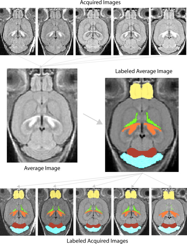
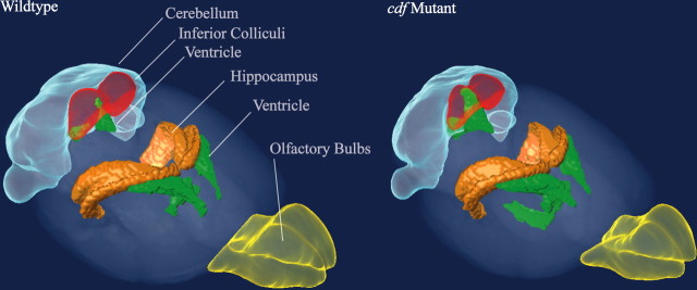
Magnetic resonance imaging and computer image analysis in human clinical studies effectively identify abnormal neuroanatomy in disease populations. As more mouse models of neurological disorders are discovered, such an approach may prove useful for translational studies. Here, we demonstrate the effectiveness of a similar strategy for mouse neuroscience studies by phenotyping mice with the cerebellar deficient folia (cdf) mutation. Using in vivo multiple-mouse magnetic resonance imaging for increased throughput, we imaged groups of cdf mutant, heterozygous, and wild-type mice and made an atlas-based segmentation of the structures in 15 individual brains. We then performed computer automated volume measurements on the structures. We found a reduced cerebellar volume in the cdf mutants, which was expected, but we also found a new phenotype in the inferior colliculus and the olfactory bulbs. Subsequent local histology revealed additional cytoarchitectural abnormalities in the olfactory bulbs. This demonstrates the utility of anatomical magnetic resonance imaging and semiautomated image analysis for detecting abnormal neuroarchitecture in mutant mice.
Figures


References
-
- Ahrens ET, Dubowitz DJ (2001). Peripheral somatosensory fMRI in mouse at 11.7 T. NMR Biomed 14:318–324. - PubMed
-
- Beierbach E, Park C, Ackerman SL, Goldowitz D, Hawkes R (2001). Abnormal dispersion of a purkinje cell subset in the mouse mutant cerebellar deficient folia (cdf). J Comp Neurol 436:42–51. - PubMed
-
- Bock NA, Nieman BJ, Bishop JB, Mark Henkelman R (2005). In vivo multiple-mouse MRI at 7 Tesla. Magn Reson Med 54:1311–1316. - PubMed
-
- Bucan M, Abel T (2002). The mouse: genetics meets behaviour. Nat Rev Genet 3:114–123. - PubMed
-
- Chen XJ, Kovacevic N, Lobaugh NJ, Sled JG, Henkelman RM, Henderson JT (2006). Neuroanatomical differences between mouse strains as shown by high-resolution 3D MRI. NeuroImage 29:99–105. - PubMed
Publication types
MeSH terms
Substances
Grants and funding
LinkOut - more resources
Full Text Sources
Medical
Molecular Biology Databases
Research Materials