. 2006 Apr 26;26(17):4460-4.
doi: 10.1523/JNEUROSCI.4935-05.2006.

Radial migration of superficial layer cortical neurons controlled by novel Ig cell adhesion molecule MDGA1

Affiliations

Radial migration of superficial layer cortical neurons controlled by novel Ig cell adhesion molecule MDGA1

Akihide Takeuchi et al. J Neurosci. .

Abstract

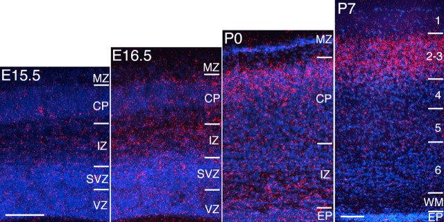
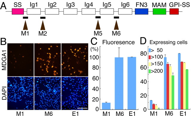
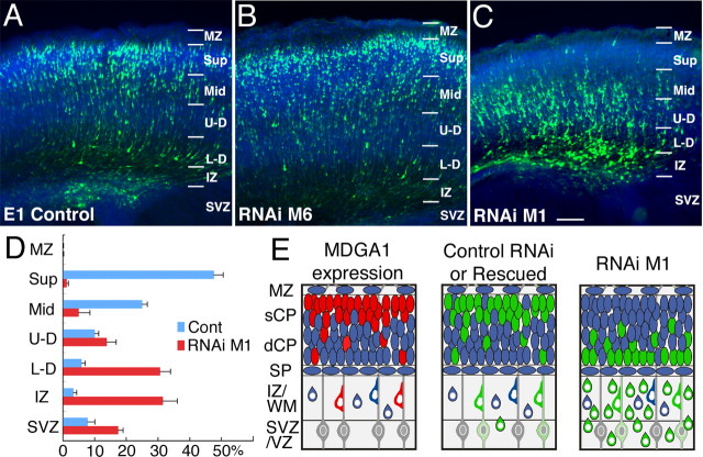
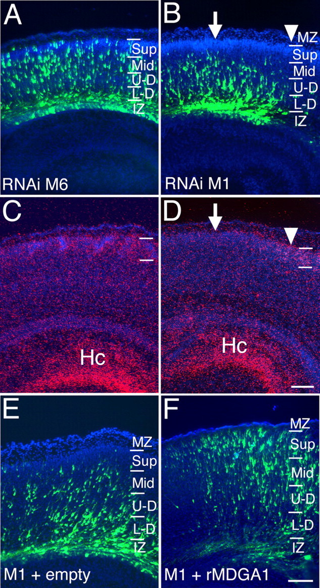
MAM (meprin/A5 protein/receptor protein tyrosine phosphatase mu) domain glycosylphosphatidylinositol anchor 1 (MDGA1), a unique cell surface glycoprotein, is similar to Ig-containing cell adhesion molecules that influence neuronal migration and process outgrowth. We show in postnatal mice that MDGA1 is expressed by layer 2/3 neurons throughout the neocortex. During development, MDGA1 is expressed in patterns consistent with its expression by migrating layer 2/3 neurons, suggesting a role for MDGA1 in controlling their migration and settling in the superficial cortical plate. To test this hypothesis, we performed loss-of-function studies using RNA interference (RNAi) targeting different sequences of mouse MDGA1. RNAi or empty vectors were coelectroporated with an enhanced green fluorescent protein reporter in utero into the lateral ventricle at embryonic day 15.5 to transfect progenitors of superficial layer neurons; the distributions of transfected neurons were analyzed late on postnatal day 0. We found a direct correlation between effectiveness of an RNAi in suppressing MDGA1 expression and disrupting migration of superficial layer neurons. An RNAi with no effect on MDGA1 expression has no effect on the migration. In contrast, an RNAi that suppresses MDGA1 expression also blocks proper migration of transfected superficial layer neurons, with essentially all transfected cells found deep in the cortical plate or beneath it. This migration defect is rescued by cotransfection of a rat MDGA1 expression construct along with the effective RNAi, confirming that the RNAi effect is specific to diminishing mouse MDGA1 expression. RNAi transfections of deep layer neurons that do not express MDGA1 do not significantly affect their migration. We conclude that MDGA1 acts cell autonomously to control the migration of MDGA1-expressing superficial layer cortical neurons.


Figures

Figure 1.
Figure 1.
Laminar expression of MDGA1 in the developing cortex. In situ hybridization of MDGA1 expression in the developing mouse cortex is shown. At E16.5, MDGA1 is modestly expressed in the IZ. At P0, expression is robust in the CP, and dense clusters of silver grains (red) deeper in the CP and IZ suggest MDGA1 expression by migrating neurons. By P7, robust expression is localized to layers 2/3. EP, Ependymal layer; WM, white matter. Scale bars, 100 μm; bar in E15.5 is E15.5 to P0.
Figure 2.
Figure 2.
Targeted sequences for RNAi vectors differentially suppress MDGA1 protein expression in vitro. A, MDGA1 domain structure and selected target regions to make RNAi vectors; the four target regions (M1, M2, M5, M6) were 19 bp sequences located between the Ig domains. SS, Signal peptide sequence; FN3, fibronectin type 3 repeat; MAM, MAM domain; GPI, glycosylphosphatidylinositol anchor. B, COS-7 cells cotransfected with a pcDNA3.1 vector containing MDGA1 cDNA tagged with a Myc epitope and an RNAi pSUPER vector or en empty pSUPER vector (E1); RNAi vector name (M1, M6) denotes sequence location shown in A. Shown is immunofluorescence using a Myc antibody 24 h after transfection; the secondary antibody was conjugated to Alexa568 (orange). Cultures are DAPI stained (blue). Scale bar, 100 μm C, Histogram of fluorescence intensity of MDGA1-Myc protein. D, Histogram of the number of MDGA1-positive cells with a total pixel intensity above the indicated thresholds. Quantitative data for M2 is statistically indistinguishable from that shown in C and D for M1 and M5 from that shown for M6.
Figure 3.
Figure 3.
Transfection of M1 RNAi to suppress MDGA1 selectively results in aberrant deep distribution of superficial layer neurons. Distribution of GFP-labeled neurons late on P0 following cotransfections on E15.5 with a GFP and E1 vector (A), GFP and M6 RNAi vector (B), and GFP and M1 RNAi vector (C). D, Five M1 cases and five control cases (4 E1 and 1 M6) were quantitatively analyzed. The cortical wall was subdivided as indicated, and numbers of GFP-labeled transfected cells in each layer were counted and expressed as a percentage of the total. Statistical analyses comparing the number of labeled cells in each layer between control and M1 transfected cases (ANOVA, Bonferroni’s test) are shown. Statistical difference is seen for each set of layers (p < 0.01) except MZ and U-D layers. Scale bars, 100 μm. E, Schematics of late P0 cortex showing an expression pattern of MDGA1 and distribution of GFP-labeled neurons transfected with M1 RNAi or control (M6 RNAi, E1) vectors at E15.5. Expression of MDGA1 is similar to distribution of GFP-labeled neurons transfected with control vectors or in rescue experiments (cotransfections with M1 RNAi and rat MDGA1 cDNA vectors). However, distribution of GFP-labeled neurons transfected with an M1 RNAi vector is aberrantly displaced deep. sCP, Superficial cortical plate; dCP, deep cortical plate; Sup, superficial CP; Mid, middle CP; U-D, upper-deep CP; L-D, lower-deep CP; others are as in Figure 1.
Figure 4.
Figure 4.
Differential suppression of MDGA1 expression in vivo by mouse RNAi vectors with rescue of RNAi-induced migration defect by cotransfection of rat MDGA1. A–D, Distribution of GFP-labeled neurons late on P0 following cotransfection with GFP and M6 (A) or GFP and M1 RNAi vectors at E15.5 (B). In situ hybridization using S35-labeled riboprobes for MDGA1 (C, D) on adjacent sections of A and B, respectively. M6 RNAi transfection has no detectable effect on MDGA1 expression. However, MDGA1 expression is diminished in superficial layers above the domain of M1 RNAi transfection (arrow) but not adjacent to it (arrowhead). E, F, Distribution of radially migrating neurons cotransfected with M1 RNAi plus empty pCMV plus GFP vectors (control) (E) and M1 RNAi plus pCMV rat-MDGA1 (rMDGA1) plus GFP vectors (F). M1 RNAi target sequence (GGAGGATAACATCAGCGAG) and rat MDGA1 cDNA (-GGAGGATAATATCAGCGAG-) have a one nucleotide mismatch (bolded). In control cases (E), most GFP-labeled cells are deep, and virtually none are in the superficial CP. As shown in F, cotransfection with rat MDGA1 expression vector essentially corrects the migration defect and shifts distribution of GFP-labeled cells compared with D, with many in superficial CP. Scale bars, 100 μm. Hc, Hippocampal formation; other abbreviations are as in Figure 1.

References

    1. Adams NC, Tomoda T, Cooper M, Dietz G, Hatten ME (2002). Mice that lack astrotactin have slowed neuronal migration. Development 129:965–972. - PubMed
    1. Anton ES, Kreidberg JA, Rakic P (1999). Distinct functions of alpha3 and alpha(v) integrin receptors in neuronal migration and laminar organization of the cerebral cortex. Neuron 22:277–289. - PubMed
    1. Bai J, Ramos RL, Ackman JB, Thomas AM, Lee RV, LoTurco JJ (2003). RNAi reveals doublecortin is required for radial migration in rat neocortex. Nat Neurosci 6:1277–1283. - PubMed
    1. Bayer S, Altman J (1991). In: Neocortical development New York: Raven.
    1. Bielas S, Higginbotham H, Koizumi H, Tanaka T, Gleeson JG (2004). Cortical neuronal migration mutants suggest separate but intersecting pathways. Annu Rev Cell Dev Biol 20:593–618. - PubMed

Publication types