. 2006 Apr 26;26(17):4546-58.
doi: 10.1523/JNEUROSCI.5412-05.2006.

Hippocampal lesion prevents spatial relational learning in adult macaque monkeys

Affiliations

Hippocampal lesion prevents spatial relational learning in adult macaque monkeys

Pamela Banta Lavenex et al. J Neurosci. .

Abstract

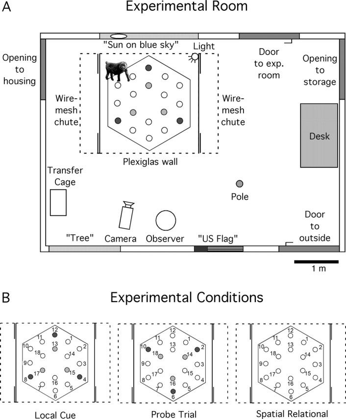
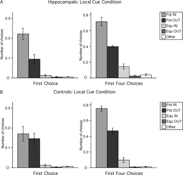
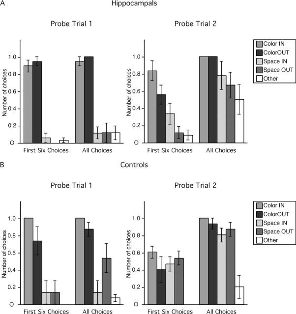
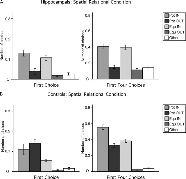
The role of the hippocampus in spatial learning and memory has been extensively studied in rodents. Comparable studies in nonhuman primates, however, are few, and findings are often contradictory. This may be attributable to the failure to distinguish between allocentric and egocentric spatial representations in experimental designs. For this experiment, six adult monkeys received bilateral hippocampal ibotenic acid lesions, and six control subjects underwent sham surgery. Freely moving monkeys then foraged for food located in two arrays of three distinct locations among 18 locations distributed in an open-field arena. Multiple goals and four pseudorandomly chosen entrance points precluded the monkeys' ability to rely on an egocentric strategy to identify food locations. Monkeys were tested in two conditions. First, local visual cues marked the food locations. Second, no local cues marked the food locations, so that monkeys had to rely on an allocentric (spatial relational) representation of the environment to discriminate these locations. Both hippocampal-lesioned and control monkeys discriminated the food locations in the presence of local cues. However, in the absence of local cues, control subjects discriminated the food locations, whereas hippocampal-lesioned monkeys were unable to do so. Interestingly, histological analysis of the brain of one control monkey whose behavior was identical to that of the experimentally lesioned animals revealed a bilateral ischemic lesion restricted to the hippocampus. These findings demonstrate that the adult monkey hippocampal formation is critical for the establishment or use of allocentric spatial representations and that selective damage of the hippocampus prevents spatial relational learning in adult nonhuman primates.


Figures

Figure 1.
Figure 1.
Schematic representation of the testing environment and experimental conditions. A, Aerial view of the experimental room. Eighteen plastic cups were regularly distributed on a hexagonal board (210 cm in diameter) placed in a square testing arena (220 cm W × 220 cm D × 220 cm H). Remotely operated sliding doors at each corner of the arena (double solid lines) allowed the animals to go in and out of the arena from wire-mesh chutes located along both sides. The front panel, the roof, and the top half of the back panel (dashed lines) were made of Plexiglas, allowing clear view of distant environmental cues; two opaque side panels (solid lines) provided visual barriers between the open-field arena and the wire-mesh holding chutes. The ability of monkeys to rely on an egocentric representation of space was precluded by alternating pseudorandomly between four different entrances into the open-field arena and using multiple goal locations. B, Schematic representation of the arena in the different testing conditions. (1) Local cue condition: blue cups marked potentially-baited locations 4, 8, 12 on the outer array, and red cups marked potentially-baited locations 13, 15, 17 on the inner array. All other locations were covered with neutral (beige) cups. (2) Probe trial: colored cups were shifted 60° from the correct spatial locations. Blue cups were at locations 2, 6, 10, and red cups were at locations 14, 16, 18. Neutral cups were at locations 4, 8, 12 and 13, 15, 17, as well as at locations 1, 3, 5, 7, 9, 11. No food was present. (3) Spatial relational condition: neutral cups covered the potentially-baited locations 4, 8, 12 and 13, 15, 17, as well as all other locations.
Figure 2.
Figure 2.
Hippocampal-lesioned (A) and control (B) monkeys' strategy in the local cue condition. Pot IN, Potentially-baited locations at the corners of the inner hexagon (locations 13, 15, 17); Pot OUT, potentially-baited locations at the corners of the outer hexagon (locations 4, 8, 12); Equ IN, never-baited locations at the corners of the inner hexagon (locations 14, 16, 18); Equ OUT, never-baited locations at the corners of the outer hexagon (locations 2, 6, 10); Other, never-baited locations on the sides of the outer hexagon (locations 1, 3, 5, 7, 9, 11). The number of choices in each category (n) is normalized according to the probability of making that choice (n of 3 for Pot IN, Pot OUT, Equ IN, and Equ OUT; n of 6 for Other).
Figure 3.
Figure 3.
Hippocampal-lesioned (A) and control (B) monkeys' choices in the two dissociation probe trials (no food present). Color IN, Red cups at never-baited locations at the corners of the inner hexagon (locations 14, 16, 18); Color OUT, blue cups at never-baited locations at the corners of the outer hexagon (locations 2, 6, 10); Space IN, neutral cups at correct spatial locations at the corners of the inner hexagon (locations 13, 15, 17); Space OUT, neutral cups at correct spatial locations at the corners of the outer hexagon (locations 4, 8, 10); Other, neutral cups at never-baited locations on the sides of the outer hexagon (locations 1, 3, 5, 7, 9, 11). The number of choices in each category (n) is normalized according to the probability of making that choice (n of 3 for Color IN, Color OUT, Space IN, and Space OUT; n of 6 for Other).
Figure 4.
Figure 4.
Hippocampal-lesioned (A) and control (B) monkeys' strategy in the spatial relational condition. For abbreviations, see Figure 2.
Figure 5.
Figure 5.
Photomicrographs illustrating the extent of the lesioned area in each experimentally lesioned monkey at four different levels along the rostrocaudal extent of the hippocampal formation. A–D, MMU26544; E–H, MMU26669; I–L, MMU28086; M–P, MMU27645; Q–T, MMU26149; U–X, MMU26296; Y–AB, nonlesioned monkey. Scale bar, 1 mm (applies to all panels).
Figure 6.
Figure 6.
Photomicrographs illustrating the extent of the lesioned area (arrows) on the left side of the brain in the control monkey, MMU26736, whose behavior was identical to that of experimentally lesioned monkeys. A–C, Rostral area exhibiting neuronal damage, gliosis, and neuropil disorganization. D–M, Caudal area exhibiting neuronal damage, gliosis, and neuropil disorganization. Individual panels represent adjacent sections separated by 240 μm. All Nissl-stained sections on which signs of damage were visible are presented. There was an area of ∼1.44 mm between C and D, with no visible neuron loss, gliosis, or disorganization of the neuropil. Scale bar, 250 μm (applies to all panels).
Figure 7.
Figure 7.
Photomicrographs illustrating the extent of the lesioned area (arrows) on the right side of the brain in the control monkey, MMU26736, whose behavior was identical to that of experimentally lesioned monkeys. A–J, Rostral area exhibiting neuronal damage, gliosis, and neuropil disorganization. K–M, Caudal area exhibiting neuronal damage, gliosis and neuropil disorganization. Individual panels represent adjacent sections separated by 240 μm. All Nissl-stained sections on which signs of damage were visible are presented. There was an area of ∼4.08 mm between J and K, with no visible neuron loss, gliosis or disorganization of the neuropil. Scale bar, 250 μm (applies to all panels).

References

    1. Alvarado MC, Bachevalier J (2005). Selective neurotoxic damage to the hippocampal formation impairs performance of the transverse patterning and location memory tasks in rhesus macaques. Hippocampus 15:118–131. - PubMed
    1. Amaral DG, Lavenex P (2006). Hippocampal neuroanatomy. In: The hippocampus book (Amaral DG, Andersen P, Bliss T, Morris RGM, O'Keefe J, eds) in press Oxford UP: Oxford.
    1. Angeli SJ, Murray EA, Mishkin M (1993). Hippocampectomized monkeys can remember one place but not two. Neuropsychologia 31:1021–1030. - PubMed
    1. Astur RS, Taylor LB, Mamelak AN, Philpott L, Sutherland RJ (2002). Humans with hippocampus damage display severe spatial memory impairments in a virtual Morris water task. Behav Brain Res 132:77–84. - PubMed
    1. Beason-Held LL, Rosene DL, Killiany RJ, Moss MB (1999). Hippocampal formation lesions produce memory impairment in the rhesus monkey. Hippocampus 9:562–574. - PubMed

Publication types

LinkOut - more resources