Beta-amyloid stimulates murine postnatal and adult microglia cultures in a unique manner
- PMID: 16641245
- PMCID: PMC6674057
- DOI: 10.1523/JNEUROSCI.4822-05.2006
Beta-amyloid stimulates murine postnatal and adult microglia cultures in a unique manner
Abstract
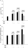
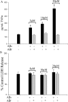
Reactive microglia are commonly observed in association with the beta-amyloid (Abeta) plaques of Alzheimer's disease brains. This localization supports the hypothesis that Abeta is a specific activating stimulus for microglia. A variety of in vitro studies have used postnatal derived rodent microglia cultures to characterize the ability of Abeta to stimulate these cells. However, it is unclear whether this paradigm accurately models conditions in aged animals. To determine whether Abeta stimulatory phenotypes differ between young and adult microglia, we established cultures of acutely isolated adult murine cortical microglia to compare with postnatal derived microglial cultures. Although cells from both ages expressed robust immunoreactivity for CD68 and CD11b, their responses to activating stimuli differed. Fibrillar Abeta was rapidly phagocytosed by postnatal microglia and both oligomeric and fibrillar peptide stimulated increased tumor necrosis factor alpha (TNFalpha) secretion. However, Abeta oligomers but not fibrils stimulated TNFalpha secretion from adult microglia. More importantly, adult microglia had diminished ability to phagocytose Abeta fibrils. These findings demonstrate that adult microglia respond to Abeta fibril stimulation uniquely from postnatal cells and suggest that adult rather than postnatal microglia cultures are more appropriate for modeling proinflammatory changes in the aged CNS.
Figures


Similar articles
-
Microglia demonstrate age-dependent interaction with amyloid-β fibrils.J Alzheimers Dis. 2011;25(2):279-93. doi: 10.3233/JAD-2011-101014. J Alzheimers Dis. 2011. PMID: 21403390 Free PMC article.
-
Inhibition of Src kinase activity attenuates amyloid associated microgliosis in a murine model of Alzheimer's disease.J Neuroinflammation. 2012 Jul 2;9:117. doi: 10.1186/1742-2094-9-117. J Neuroinflammation. 2012. PMID: 22673542 Free PMC article.
-
Isolated amyloid-β(1-42) protofibrils, but not isolated fibrils, are robust stimulators of microglia.ACS Chem Neurosci. 2012 Apr 18;3(4):302-11. doi: 10.1021/cn2001238. Epub 2012 Jan 9. ACS Chem Neurosci. 2012. PMID: 22860196 Free PMC article.
-
Impact of prenatal immune system disturbances on brain development.J Neuroimmune Pharmacol. 2013 Mar;8(1):79-86. doi: 10.1007/s11481-012-9374-z. Epub 2012 May 13. J Neuroimmune Pharmacol. 2013. PMID: 22580757 Review.
-
Aging, microglial cell priming, and the discordant central inflammatory response to signals from the peripheral immune system.J Leukoc Biol. 2008 Oct;84(4):932-9. doi: 10.1189/jlb.0208108. J Leukoc Biol. 2008. PMID: 18495785 Free PMC article. Review.
Cited by
-
Amyloid precursor protein and proinflammatory changes are regulated in brain and adipose tissue in a murine model of high fat diet-induced obesity.PLoS One. 2012;7(1):e30378. doi: 10.1371/journal.pone.0030378. Epub 2012 Jan 19. PLoS One. 2012. PMID: 22276186 Free PMC article.
-
Various Cell Populations Within the Mononuclear Fraction of Bone Marrow Contribute to the Beneficial Effects of Autologous Bone Marrow Cell Therapy in a Rodent Stroke Model.Transl Stroke Res. 2016 Aug;7(4):322-30. doi: 10.1007/s12975-016-0462-x. Epub 2016 Mar 20. Transl Stroke Res. 2016. PMID: 26997513 Free PMC article.
-
Characterization of Novel Src Family Kinase Inhibitors to Attenuate Microgliosis.PLoS One. 2015 Jul 10;10(7):e0132604. doi: 10.1371/journal.pone.0132604. eCollection 2015. PLoS One. 2015. PMID: 26161952 Free PMC article.
-
Animal Models of Alzheimer's Disease: Utilization of Transgenic Alzheimer's Disease Models in Studies of Amyloid Beta Clearance.Curr Transl Geriatr Exp Gerontol Rep. 2012 Mar;1(1):11-20. doi: 10.1007/s13670-011-0004-z. Epub 2012 Jan 19. Curr Transl Geriatr Exp Gerontol Rep. 2012. PMID: 23440676 Free PMC article.
-
Brain metabolic stress and neuroinflammation at the basis of cognitive impairment in Alzheimer's disease.Front Aging Neurosci. 2015 May 19;7:94. doi: 10.3389/fnagi.2015.00094. eCollection 2015. Front Aging Neurosci. 2015. PMID: 26042036 Free PMC article. Review.
References
-
- Akiyama H, Barger S, Barnum S, Bradt B, Bauer J, Cole GM, Cooper NR, Eikelenboom P, Emmerling M, Fiebich BL, Finch CE, Frautschy S, Griffin WS, Hampel H, Hull M, Landreth G, Lue L, Mrak R, Mackenzie IR, McGeer PL (2000). Inflammation and Alzheimer's disease. Neurobiol Aging 21:383–421. - PMC - PubMed
-
- Becher B, Antel JP (1996). Comparison of phenotypic and functional properties of immediately ex vivo and cultured human adult microglia. Glia 18:1–10. - PubMed
-
- Carson MJ, Reilly CR, Sutcliffe JG, Lo D (1998). Mature microglia resemble immature antigen-presenting cells. Glia 22:72–85. - PubMed
-
- Chromy BA, Nowak RJ, Lambert MP, Viola KL, Chang L, Velasco PT, Jones BW, Fernandez SJ, Lacor PN, Horowitz P, Finch CE, Krafft GA, Klein WL (2003). Self-assembly of Abeta(1–42) into globular neurotoxins. Biochemistry 42:12749–12760. - PubMed
Publication types
MeSH terms
Substances
Grants and funding
LinkOut - more resources
Full Text Sources
Medical
Research Materials
