Comparative Study
. 2006 Apr 26;26(17):4672-80.
doi: 10.1523/JNEUROSCI.5539-05.2006.

Omega-3 fatty acids improve recovery, whereas omega-6 fatty acids worsen outcome, after spinal cord injury in the adult rat

Affiliations
Comparative Study

Omega-3 fatty acids improve recovery, whereas omega-6 fatty acids worsen outcome, after spinal cord injury in the adult rat

Von R King et al. J Neurosci. .

Abstract

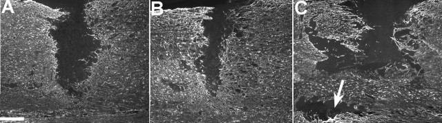
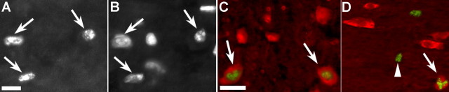
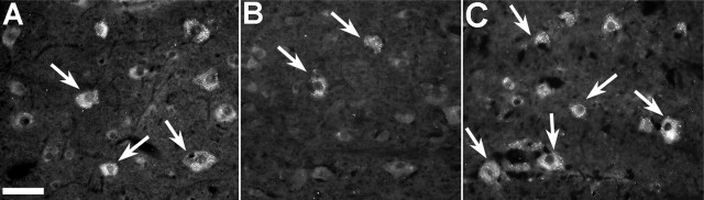
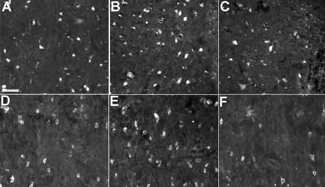
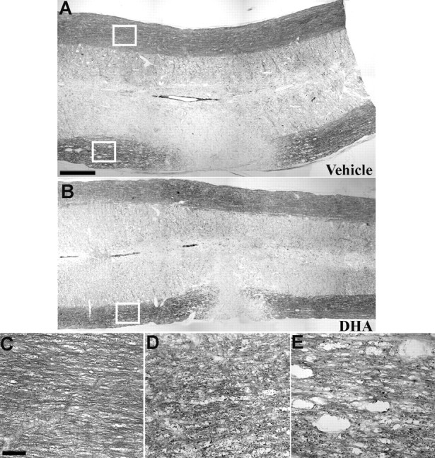
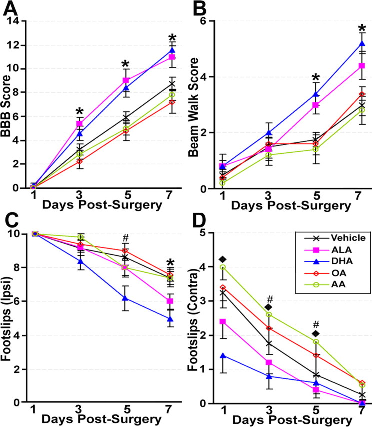
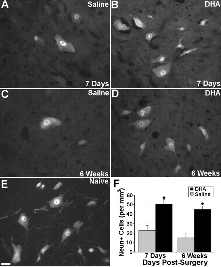
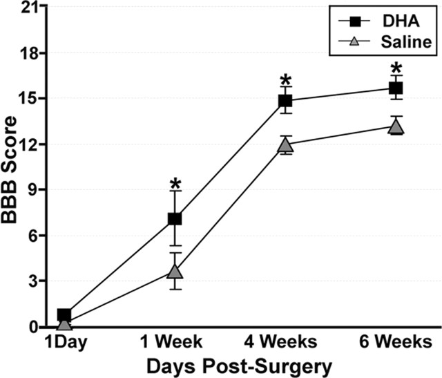
Spinal cord injury (SCI) is a cause of major neurological disability, and no satisfactory treatment is currently available. Evidence suggests that polyunsaturated fatty acids (PUFAs) could target some of the pathological mechanisms that underlie damage after SCI. We examined the effects of treatment with PUFAs after lateral spinal cord hemisection in the rat. The omega-3 PUFAs alpha-linolenic acid and docosahexaenoic acid (DHA) injected 30 min after injury induced significantly improved locomotor performance and neuroprotection, including decreased lesion size and apoptosis and increased neuronal and oligodendrocyte survival. Evidence showing a decrease in RNA/DNA oxidation suggests that the neuroprotective effect of omega-3 PUFAs involved a significant antioxidant function. In contrast, animals treated with arachidonic acid, an omega-6 PUFA, had a significantly worse outcome than controls. We confirmed the neuroprotective effect of omega-3 PUFAs by examining the effects of DHA treatment after spinal cord compression injury. Results indicated that DHA administered 30 min after spinal cord compression not only greatly increased survival of neurons but also resulted in significantly better locomotor performance for up to 6 weeks after injury. This report shows a striking difference in efficacy between the effects of treatment with omega-3 and omega-6 PUFAs on the outcome of SCI, with omega-3 PUFAs being neuroprotective and omega-6 PUFAs having a damaging effect. Given the proven clinical safety of omega-3 PUFAs, our observations show that these PUFAs have significant therapeutic potential in SCI. In contrast, the use of preparations enriched in omega-6 PUFAs after injury could worsen outcome after SCI.


Figures

Figure 1.
Figure 1.
GFAP labeling at the spinal cord hemisection site after a single intravenous injection of either saline or PUFAs after 7 d of survival. Vehicle (A) and OA-treated (not pictured) animals had similarly sized lesions that extended laterally from the midline of the spinal cord. Lesions in animals treated with DHA (B) or ALA (not pictured) were similar, with both groups having smaller lesions than either vehicle- or OA-treated animals. In contrast, AA-treated animals (C) had greatly expanded lesions, with some animals showing islands of damage discontinuous with the original lesion site (arrow). Scale bar, 200 μm.
Figure 2.
Figure 2.
TUNEL labeling of apoptotic nuclei adjacent to the spinal cord hemisection site after a single intravenous injection of either saline or PUFAs after 7 d of survival. TUNEL-labeled nuclei (A) were clearly identifiable in all treatment groups and were confirmed by counterstaining with the nuclear stain 4′,6-diamidino-2-phenylindole (B) (corresponding arrows in A and B show double-labeled nuclei). Double labeling (C) for TUNEL (green) and NeuN (red) showed occasional apoptotic nuclei within neurons (arrows). Similarly, double labeling (D) for APC (red) and TUNEL (green) showed occasional double-labeled oligodendrocytes (arrow). Numerous TUNEL single-labeled cells were also present (arrowhead). Scale bars: A–D, 50 μm.
Figure 3.
Figure 3.
oh8G labeling of RNA/DNA oxidation in cells adjacent to the spinal cord hemisection site after a single intravenous injection of either saline or PUFAs after 7 d of survival. The number of oh8G-labeled neurons in the area immediately adjacent to the lesion site in vehicle-treated (A) and OA-treated (not pictured) animals was similar. Both of these groups had noticeably more oh8G-labeled cells than either DHA-treated (B) and ALA-treated (not pictured) animals, which had few oh8G-labeled cells. In contrast, AA-treated animals (C) had substantially more oh8G-labeled cells than all other groups. Scale bar, 50 μm.
Figure 4.
Figure 4.
NeuN labeling of neurons and APC labeling of oligodendrocytes adjacent to the spinal cord hemisection site after a single intravenous injection of either saline or PUFAs after 7 d of survival. The number of NeuN-labeled neurons in the area immediately adjacent to the lesion site in vehicle treated (A) and OA-treated (not pictured) animals was similar. Both of these groups had noticeably fewer NeuN-labeled cells than DHA-treated (B) and ALA-treated (not pictured) animals, which had similar numbers of neurons. In contrast, AA-treated animals (C) had substantially fewer NeuN-labeled neurons than all other groups. Similar results were seen with APC-labeled oligodendrocytes, with vehicle-treated (D) and OA-treated (not pictured) animals having fewer cells than DHA-treated (E) and ALA-treated (not pictured) animals. AA-treated animals (F) had fewer oligodendrocytes than the other groups. Scale bar: (in A) A–F, 250 μm.
Figure 5.
Figure 5.
Luxol fast blue staining of the lateral whiter matter after spinal cord hemisection in animals that received a single intravenous injection of either saline or PUFAs after 7 d of survival. The appearance of myelin staining at the lesion site in vehicle-treated (A), DHA-treated (B), and ALA-, OA-, and AA-treated (not pictured) animals was similar, with all groups showing a lack of staining in the white matter ipsilateral to the lesion. Adjacent to the lesion site, the myelin in the white matter ipsilateral to the hemisection was disrupted relative to the uninjured side (C, enlargement of area within the top box in A) in all groups. However, myelin in ALA-treated (not pictured) and DHA-treated (D, enlargement of area within the box in B) animals was clearly less disrupted compared with OA-, AA-, and vehicle-treated animals (E, enlargement of area within the bottom box in A), which had large cavities in the white matter several millimeters from the lesion site. Scale bars: (in A) A, B, 250 μm; (in C) C–E, 25 μm.
Figure 6.
Figure 6.
Locomotor performance after spinal cord hemisection in animals that received a single intravenous injection of either saline or PUFAs. Both ALA- and DHA-treated animals performed significantly better on the BBB open field (A) and the beam walk (B) tasks than the remaining groups, which did not differ. DHA- and ALA-treated animals also had significantly fewer foot slips with the hindlimb ipsilateral to the lesion on the horizontal ladder (C) compared with the other groups, although this difference was only seen for the ALA group at 7 d after surgery. In contrast, DHA- but not ALA-treated animals showed significantly fewer slips with the hindlimb contralateral to the lesion, compared with vehicle- and OA-treated animals, whereas AA-treated animals were worse on this parameter than all of the other groups (D). *DHA- and ALA-treated animals significantly different from remaining groups (p < 0.05); #DHA-treated animals significantly different from the remaining groups (p < 0.05); ♦, vehicle-, DHA-, and ALA-treated animals significantly different from AA-treated animals (p < 0.05).
Figure 7.
Figure 7.
NeuN labeling of neurons after spinal cord compression and a single intravenous injection of either DHA or saline. At 1 week after surgery, the number of NeuN-labeled cells in saline-treated animals (A) was substantially less than naive control animals (E). DHA-treated animals (B, D) also had more NeuN-labeled neurons compared with saline-treated (A, C) animals at both 1 and 6 weeks after surgery. Quantitative analysis (F) confirmed that there were significantly more NeuN-labeled neurons in DHA-treated animals at both 1 week and 6 weeks after surgery compared with saline-treated controls. Scale bar: (in E) A–E, 50 μm. *DHA-treated animals significantly different from saline-treated controls (p < 0.05). Error bars represent SEM.
Figure 8.
Figure 8.
Locomotor performance of animals that received a spinal cord compression injury and a single intravenous injection of either DHA or saline. Both DHA- and saline-treated animals showed similar low levels of locomotor function 1 d after surgery as assessed on the BBB open field task. However, by 1 week after surgery, DHA-treated animals show significantly better locomotor function (3 points on the BBB scale) compared with saline-treated animals, a difference that was maintained until 6 weeks after surgery (the latest point examined). *DHA-treated animals significantly different from saline-treated controls (p < 0.05).

References

    1. Basso DM, Beattie MS, Bresnahan JC (1996). Graded histological and locomotor outcomes after spinal cord contusion using the NYU weight-drop device versus transection. Exp Neurol 139:244–256. - PubMed
    1. Bazan NG (2005). Neuroprotectin D1 (NPD1): a DHA-derived mediator that protects brain and retina against cell injury-induced oxidative stress. Brain Pathol 15:159–166. - PMC - PubMed
    1. Beattie MS, Farooqui AA, Bresnahan JC (2000). Rev of current evidence for apoptosis after spinal cord injury. J Neurotrauma 17:915–925. - PubMed
    1. Blondeau N, Widmann C, Lazdunski M, Heurteaux C (2002). Polyunsaturated fatty acids induce ischemic and epileptic tolerance. Neuroscience 109:231–241. - PubMed
    1. Calder PC (2003). Long-chain n-3 fatty acids and inflammation: potential application in surgical and trauma patients. Braz J Med Biol Res 36:433–446. - PubMed

Publication types

MeSH terms