. 2006 May;80(10):4729-39.
doi: 10.1128/JVI.80.10.4729-4739.2006.

NS1 interaction with CKII alpha: novel protein complex mediating parvovirus-induced cytotoxicity

Affiliations

NS1 interaction with CKII alpha: novel protein complex mediating parvovirus-induced cytotoxicity

Jürg P F Nüesch et al. J Virol. 2006 May.

Abstract

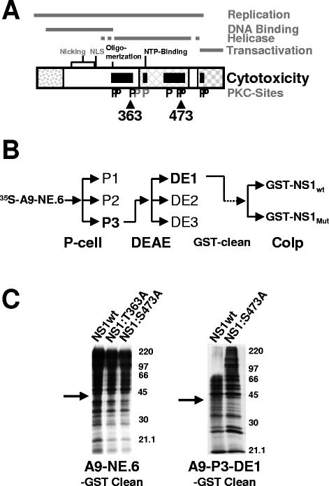
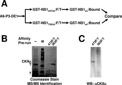
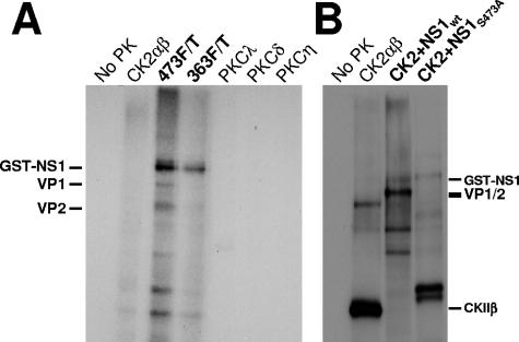
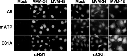
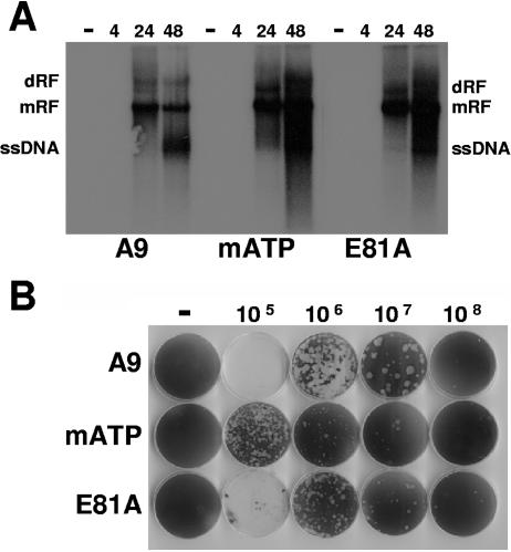
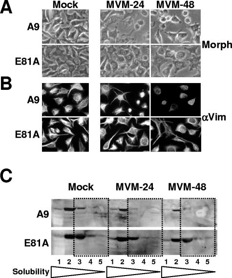
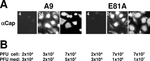
During a productive infection, the prototype strain of the parvovirus minute virus of mice (MVMp) induces dramatic morphological alterations in permissive A9 fibroblasts, culminating in cell lysis at the end of infection. These cytopathic effects (CPE) result from rearrangements and destruction of the cytoskeletal micro- and intermediate filaments, while other structures such as the nuclear lamina and particularly the microtubule network remain protected throughout the infection (J. P. F. Nüesch et al., Virology 331:159-174, 2005). In order to unravel the mechanism(s) by which parvoviruses trigger CPE, we searched for NS1 interaction partners by differential affinity chromatography, using distinct NS1 mutants debilitated specifically for this function. Thereby, we isolated an NS1 partner polypeptide, whose interaction with NS1 correlated with the competence of the viral product for CPE induction, and further identified it by tandem mass spectrometry and Western blotting analyses to consist of the catalytic subunit of casein kinase II, CKIIalpha. This interaction of NS1 with CKIIalpha suggested interference by the viral protein with intracellular signaling. Using permanent cell lines expressing dominant-negative CKIIalpha mutants, we were able to show that this kinase activity was indeed specifically involved in parvoviral CPE and progeny particle release. Furthermore, the NS1/CKIIalpha complex proved to be able to specifically phosphorylate viral capsids, indicating a mediator function of NS1 for CKII activity and specificity, at least in vitro. Altogether our data suggest that parvovirus-induced CPE is mediated by NS1 interference with intracellular CKII signaling.


Figures

FIG. 1.
FIG. 1.
Cellular interaction partners of NS1 lacking affinity to cytotoxicity mutants. (A) Domain structure of NS1 (25). The common N terminus of NS1 and NS2 is shown as a dotted box. The crosshatched box represents the homology region with SV40 large-T antigen, and the checkered box denotes the transactivation domain. NS1 domains involved in the DNA binding, helicase, and overall replication functions of the viral product are delineated on top. The nucleotide-binding site (NTP-Binding), oligomerization region, nuclear localization signal (NLS), and DNA-nicking motifs (metal coordination site and linkage tyrosine) are indicated. NS1 regions harboring mutations affecting CPE induction are represented as black boxes. Consensus PKC phosphorylation sites (P) are indicated in black if the corresponding mutant was impaired in its ability to induce CPE and in gray if no significant change was detected compared to the wild-type protein. Positions of NS1 mutants T363 and S473 used to search for NS1 interaction partners are indicated. (B) Cell fractionation procedure used to enrich for “rare” NS1 interaction partners. P-cell, phosphocellulose column with P1, flow-through at 150 mM NaCl concentration; P2, eluate at 400 mM NaCl; P3, eluate at 1 M NaCl. DEAE, DE52 anion-exchange column with DE1, flow-through at 200 mM NaCl; DE2, eluate at 400 mM NaCl; DE3, eluate at 1 M NaCl. Cellular proteins in P3-DE1 with affinity for GST were purified on GST-coated glutathione Sepharose beads; residual polypeptides were then coimmunoprecipitated with either wild-type or mutant GST-tagged NS1. (C) Coimmunoprecipitation of 35S-labeled cellular proteins with GST-NS1 derived from vaccinia virus expression in HeLa cells. The left panel shows a comparison between wild-type NS1, NS1:T363A, and NS1:S473A using total nuclear squeeze from A9 cells, while the right panel presents a comparison between wild-type NS1 and NS1:S473A using partially purified proteins after phosphocellulose and DE52 column chromatography. Proteins with affinity for the GST tag have been eliminated prior to the immunoprecipitation with GST-NS1. The arrow points at a ∼40-kDa polypeptide specifically interacting with wild-type NS1, while lacking affinity to the cytotoxic NS1 mutant S473A.
FIG. 2.
FIG. 2.
Isolation and identification of cellular proteins interacting with wild-type but not nontoxic mutant NS1. (A) Purification of cytotoxicity-related NS1 partner proteins by sequential NS1 affinity chromatography. The P3-DE1 prepurified protein fractions were run first over a column carrying either cytotoxicity mutant of NS1 (T363A or S473A). Unbound proteins (flow-through; F/T) failing to interact with the nontoxic NS1 derivative were then run over a second affinity column coupled with wild-type GST-NS1 to capture cytotoxicity-dependent NS1 partners. After extensive washing, eluates were collected, containing the cellular proteins which bind to wild-type but not cytotoxic mutant NS1 proteins. (B) SDS-PAGE separation and Coomassie blue staining of proteins retained by GST-NS1wt affinity columns and subsequently eluted at 700 mM NaCl. A9 cell proteins from the P3-DE1 fraction (see Fig. 1B) were run over the GST-NS1wt column without prior additional purification (−, total NS1-binding protein) or after prior elimination of proteins binding to either mutant NS1 affinity column (473F/T; proteins lacking affinity to GST-NS1:S473A and binding to wild-type NS1; 363F/T, proteins lacking affinity to GST-NS1:T363A and binding to wild-type NS1). NS1wt partner proteins overrepresented in 474F/T versus 363F/T, and conversely, are marked with dots. Marked bands were cut from the gel and submitted to MS/MS analyses. The 40-kDa species overrepresented in 473F/T (i.e., with a reduced affinity for NS1:S473A) was identified as CKIIα. M, molecular weight markers (Amersham-Pharmacia). (C) This identification was confirmed by Western blotting (WB) of the two protein fractions obtained after differential affinity chromatography with NS1 variants using antibodies specific for CKIIα.
FIG. 3.
FIG. 3.
In vitro kinase activity of the NS1/CKIIα complex. (A) CsCl-purified MVMp capsids were subjected to phosphorylation using [γ-32P]ATP and indicated recombinant protein kinases or the NS1wt complexes with 473F/T or 363F/T proteins. Treated capsids were immunoprecipitated with the monoclonal antibody EIIF3 and analyzed by 10% SDS-PAGE and autoradiography. The migrations of GST-NS1 (∼110 kDa), VP1, and VP2 are indicated. (B) Phosphorylation of MVMp capsids with recombinant CKIIαβ either alone or in the presence of wild-type or mutant (S473A) GST-tagged NS1 proteins produced by recombinant vaccinia virus expression. Labeled proteins were analyzed directly by 12% SDS-PAGE and autoradiography. The migration of GST-NS1, VP1, and VP2, as well as the regulatory subunit of CKII, CKIIβ (which is a target for CKII phosphorylation), is indicated.
FIG. 4.
FIG. 4.
Nucleotide and amino acid sequence of mouse CKIIα cDNA derived from A9 cells. The cDNA was isolated from an A9 cDNA bank using the primers indicated by arrows. Differences between the nucleotide sequence obtained and the published rat CKIIα sequence (NCBI L15618) are indicated in boldface and italics, with corresponding amino acid changes shown in boldface and by the triple-letter code. Amino acids targeted for mutagenesis to generate dominant-negative mutants are indicated by hatched boxes, while the nuclear targeting sequence is shown in light gray.
FIG. 5.
FIG. 5.
Immunofluorescence detection of CKIIα expressed from the endogenous gene and transfected mutant clones. A9 cells or derivatives thereof stably transfected with indicated mutants of CKIIα expression constructs were grown on spot slides, infected (or not) with MVMp (30 PFU/cell), and fixed 24 or 48 h p.i. CKIIα was detected using a polyclonal goat antiserum (αCKII) and FITC-conjugated anti-goat IgGs. NS1 was revealed using a polyclonal rabbit anti-NS1C antiserum (αNS1) and rhodamine-conjugated anti-rabbit IgGs.
FIG. 6.
FIG. 6.
MVMp replication in A9 cells and derivatives expressing dominant-negative CKIIα. (A) Asynchronously growing A9 cell derivatives (A9-P38:CKIImATP and A9-P38:CKIIE81A) expressing CKIIα variants under control of the viral P38 promoter were infected (or not) with MVMp (30 PFU/cell). The accumulation of double-stranded replicative forms and single-stranded virion DNA (ssDNA) was determined by Southern blotting at 4 h, 24 h, and 48 h p.i. dRF, dimer replicative form; mRF, monomer replicative form. Prolonged exposure showed similar amounts of input ssDNA in infected A9 cells and derivatives at 4 h p.i. (data not shown), indicating that modulation of the casein kinase II pathway did not affect virus entry. (B) Determination of MVMp plaquing efficiency and plaque morphology in A9 cells and derivatives expressing dominant-negative CKIIα according to standard plaque assays.
FIG. 7.
FIG. 7.
MVMp-induced CPE in the presence of a dominant-negative CKIIα mutants (E81A). (A) Asynchronously growing A9 cells were infected (or not) with MVMp (30 PFU/cell) and examined for morphological alterations (Morph) by phase-contrast microscopy. (B) Asynchronously growing A9 cells grown on spot slides were infected (or not) with MVMp (30 PFU/cell) and fixed with paraformaldehyde at indicated times postinfection. Preparations were analyzed by immunofluorescence for the organization of vimentin filaments using goat polyclonal antiserum raised against vimentin (αVim) and FITC-coupled anti-goat IgGs. (C) Biochemical determination of MVMp-induced alterations to the cytoskeleton. Asynchronously growing A9 cells were infected with MVMp (30 PFU/cell), harvested 24 h and 48 h p.i., and analyzed for the solubility of the intermediate filament vimentin. Cell extracts were treated with increasing amounts and strengths of detergents, and the individual fractions (S1 to S5) were analyzed for their vimentin content by Western blotting. The most insoluble filament proteins are indicated by dotted frames.
FIG. 8.
FIG. 8.
Release of progeny virus particles from A9 cells and derivatives expressing dominant-negative CKIIα mutants. (A) Cells grown on spot slides were fixed with paraformaldehyde at 4 h, 24 h, and 48 h p.i. and analyzed by immunofluorescence using a mixture of two mouse monoclonal anticapsid (αCap) antibodies (EIIF3 and B7) and Cy2-conjugated anti-mouse IgGs. (B) Cell-associated infectious virions (PFU cell) and progeny particles released into the medium (PFU med) were determined by standard plaque assays.

References

    1. Anouja, F., R. Wattiez, S. Mousset, and P. Caillet-Fauquet. 1997. The cytotoxicity of the parvovirus minute virus of mice nonstructural protein NS1 is related to changes in the synthesis and phosphorylation of cell proteins. J. Virol. 71:4671-4678. - PMC - PubMed
    1. Bashir, T., J. Rommelaere, and C. Cziepluch. 2001. In vivo accumulation of cyclin A and cellular replication factors in autonomous parvovirus minute virus of mice-associated replication bodies. J. Virol. 75:4394-4398. - PMC - PubMed
    1. Caillet-Fauquet, P., M. Perros, A. Brandenburger, P. Spegelaere, and J. Rommelaere. 1990. Programmed killing of human cells by means of an inducible clone of parvoviral genes encoding non-structural proteins. EMBO J. 9:2989-2995. - PMC - PubMed
    1. Christensen, J., S. F. Cotmore, and P. Tattersall. 1997. Parvovirus initiation factor PIF: a novel human DNA-binding factor which coordinately recognizes two ACGT motifs. J. Virol. 71:5733-5741. - PMC - PubMed
    1. Christensen, J., and P. Tattersall. 2002. Parvovirus initiator protein NS1 and RPA coordinate replication fork progression in a reconstituted DNA replication system. J. Virol. 76:6518-6531. - PMC - PubMed

MeSH terms