Comparative Study
. 2006 May;80(10):4820-32.
doi: 10.1128/JVI.80.10.4820-4832.2006.

Rotavirus viremia and extraintestinal viral infection in the neonatal rat model

Affiliations
Comparative Study

Rotavirus viremia and extraintestinal viral infection in the neonatal rat model

Sue E Crawford et al. J Virol. 2006 May.

Abstract

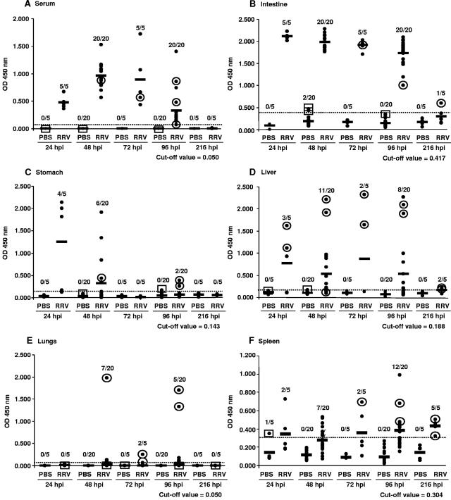
Rotaviruses infect mature, differentiated enterocytes of the small intestine and, by an unknown mechanism, escape the gastrointestinal tract and cause viremia. The neonatal rat model of rotavirus infection was used to determine the kinetics of viremia, spread, and pathology of rotavirus in extraintestinal organs. Five-day-old rat pups were inoculated intragastrically with an animal (RRV) or human (HAL1166) rotavirus or phosphate-buffered saline. Blood was collected from a subset of rat pups, and following perfusion to remove residual blood, organs were removed and homogenized to analyze rotavirus-specific antigen by enzyme-linked immunosorbent assay and infectious rotavirus by fluorescent focus assay or fixed in formalin for histology and immunohistochemistry. Viremia was detected following rotavirus infection with RRV and HAL1166. The RRV 50% antigenemia dose was 1.8 x 10(3) PFU, and the 50% diarrhea dose was 7.7 x 10(5) PFU, indicating that infection and viremia occurred in the absence of diarrhea and that detecting rotavirus antigen in the blood was a more sensitive measure of infection than diarrhea. Rotavirus antigens and infectious virus were detected in multiple organs (stomach, intestines, liver, lungs, spleen, kidneys, pancreas, thymus, and bladder). Histopathological changes due to rotavirus infection included acute inflammation of the portal tract and bile duct, microsteatosis, necrosis, and inflammatory cell infiltrates in the parenchymas of the liver and lungs. Colocalization of structural and nonstructural proteins with histopathology in the liver and lungs indicated that the histological changes observed were due to rotavirus infection and replication. Replicating rotavirus was also detected in macrophages in the lungs and blood vessels, indicating a possible mechanism of rotavirus dissemination. Extraintestinal infectious rotavirus, but not diarrhea, was observed in the presence of passively or actively acquired rotavirus-specific antibody. These findings alter the previously accepted concept of rotavirus pathogenesis to include not only gastroenteritis but also viremia, and they indicate that rotavirus could cause a broad array of systemic diseases in a number of different organs.


Figures

FIG. 1.
FIG. 1.
Kinetics of rotavirus-specific antigen detection by ELISA and infectious virus detection by FFA in PBS- or RRV-inoculated rat pups. (A) Serum; (B) intestines; (C) stomach; (D) liver; (E) lungs; (F) spleen. Each dot represents an individual rat pup in each group, and each solid horizontal line represents the average OD value of each group at the indicated time point. The numbers above each column indicate the number of ELISA positive samples over the number of samples tested. The negative cutoff value for each organ, represented by the horizontal dotted line on each graph and the indicated numeric value, was determined by calculating 3 standard deviations above the mean of the OD values of the PBS control samples. The infectivities of samples were determined either by direct FFA or by passage of the sample on MA104 cells prior to FFA. Detection (circle around the dot) or no detection (box around the dot) of infectious virus is indicated for each sample tested.
FIG. 2.
FIG. 2.
Histopathology and rotavirus antigen detection in the livers and lungs of PBS- and RRV-inoculated rat pups. Hematoxylin- and eosin-stained liver sections from RRV-inoculated rat pups at 48 hpi are shown in panels A to G. (A) Acute inflammation of the central vein (CV) and portal tract (portal vein [PV]); (B) increased magnification of panel A showing inflammation of the portal vein (PV), portal arteries (PA), and bile duct (BD); (C) microsteatosis in zone 3 (circled areas); (D) increased magnification of the boxed area of microsteatosis shown in panel C; (E) focal parenchymal necrosis; (F) serial section of the area of necrosis shown in panel E stained with anti-NSP4, with immunoperoxidase detection; (G) infiltrates of acute inflammatory cells in the necrotic parenchyma of the liver (arrows). Hematoxylin- and eosin-stained lung sections are shown in panels H and I. (H) Infiltrates of inflammatory cells in RRV-inoculated rat pups at 48 hpi; (I) normal lung morphology in PBS-inoculated rat pups at 48 hpi. Immunoperoxidase detection of rotavirus antigen is shown for serial sections of RRV-inoculated pup livers at 96 hpi stained with (J) anti-2/6-VLP and (K) anti-NSP4 antibody. The boxed area shows microsteatosis due to rotavirus infection, and the arrow indicates infiltrates of acute inflammatory cells. (L) Anti-NSP4-stained liver from PBS-inoculated pup at 48 hpi. Magnification: A, H, and I, ×50; B, ×400; C, ×100; D, ×1,000; E to G, ×250; J to L, ×10.
FIG. 3.
FIG. 3.
Detection of rotavirus antigen in lungs, hearts, thymuses, spleens, macrophages in lungs, and livers and colocalization of rotavirus antigen and macrophages in PBS- or RRV-inoculated rat pups. Immunoperoxidase detection of rotavirus antigen by anti-NSP4 antibody was done for lung sections of (A) RRV- or (B) PBS-inoculated rat pups at 96 hpi (arrows indicate morphologically identified macrophages), heart sections from (C) RRV- (arrow indicates stained cells) or (D) PBS-inoculated rat pups at 96 hpi, thymus sections from (E) RRV- or (F) PBS-inoculated rat pups at 96 hpi, spleen sections from (G) RRV- or (H) PBS-inoculated rat pups at 96 hpi, and (I) a macrophage in a blood vessel in a section of thymus from an RRV-inoculated rat pup at 96 hpi (arrow). Immunoperoxidase detection of macrophages by anti-CD68 staining was done for lung sections from (J) RRV- or (K) PBS-inoculated rat pups at 96 hpi (arrows indicate macrophages) and liver sections from (L) RRV- or (M) PBS-inoculated rat pups at 96 hpi. (N) Immunofluorescence detection of NSP4 and the macrophage marker CD68 in a lung section from an RRV-inoculated rat pup at 96 hpi, pseudocolored for NSP4 in green, for macrophages in red, and for nuclei in blue. Colocalization of NSP4 and the macrophage marker is shown in yellow. Arrows show pneumocytes infected with RRV, as detected by NSP4 staining. Magnification: A and B, ×1,000; J and K, ×300; C, D, I, and N, ×500; E, F, G, and H, ×400; L and M, ×10.
FIG. 4.
FIG. 4.
Detection of rotavirus NSP4 antigen in mouse macrophages. Confluent monolayers of mouse RAW 264.7 macrophages were infected with human rotavirus strain K8 (P3A[9], G1), DS-1 (P1B[4], G2), or YO (P1A[8], G3) or were mock infected. Replicating virus was detected using NSP4 antibody and Alexa 488 anti-rabbit IgG.
FIG. 5.
FIG. 5.
Kinetics of rotavirus-specific antigen detection by ELISA and infectious virus detection by FFA in PBS- or HAL1166-inoculated rat pups. (A) Serum; (B) liver. Each diamond represents an individual rat pup in each group, and each solid horizontal line represents the average OD value of each group at the indicated time point. The numbers above each column indicate the number of ELISA positive samples over the number of samples tested. The negative cutoff value for each organ, represented by the horizontal dotted line on each graph and the indicated numeric value, was determined by calculating 3 standard deviations above the mean of the OD values of the PBS control samples. The infectivities of samples were determined either by direct FFA or by passage of the sample on MA104 cells prior to FFA. Detection (circle around the diamond) or no detection (box around the diamond) of infectious virus is indicated for each sample tested.

References

    1. Azevedo, A. S., L. Yuan, K. I. Jeong, A. González, T. V. Nguyen, S. Pouly, M. Gochnauer, W. Zhang, A. Azevedo, and L. J. Saif. 2005. Viremia and nasal and rectal shedding of rotavirus in gnotobiotic pigs inoculated with Wa human rotavirus. J. Virol. 79:5428-5436. - PMC - PubMed
    1. Ball, J. M., P. Tian, C. Q. Zeng, A. P. Morris, and M. K. Estes. 1996. Age-dependent diarrhea induced by a rotaviral nonstructural glycoprotein. Science 272:101-104. - PubMed
    1. Barton, E. S., B. E. Youree, D. H. Ebert, J. C. Forrest, J. L. Connolly, T. Valyi-Nagy, K. Washington, J. D. Wetzel, and T. S. Dermody. 2003. Utilization of sialic acid as a coreceptor is required for reovirus-induced biliary disease. J. Clin. Investig. 111:1823-1833. - PMC - PubMed
    1. Blutt, S. E., C. D. Kirkwood, V. Parreño, K. L. Warfield, M. Ciarlet, M. K. Estes, K. Bok, R. F. Bishop, and M. E. Conner. 2003. Rotavirus antigenaemia and viraemia: a common event? Lancet 362:1445-1449. - PubMed
    1. Boshuizen, J. A., J. H. J. Reimerink, A. M. Korteland-van Male, V. J. J. van Ham, M. P. G. Koopmans, H. A. Büller, J. Dekker, and A. W. C. Einerhand. 2003. Changes in small intestinal homeostasis, morphology, and gene expression during rotavirus infection of infant mice. J. Virol. 77:13005-13016. - PMC - PubMed

Publication types

Substances