

Simian immunodeficiency virus SIVagm.sab infection of Caribbean African green monkeys: a new model for the study of SIV pathogenesis in natural hosts
- PMID: 16641277
- PMCID: PMC1472068
- DOI: 10.1128/JVI.80.10.4858-4867.2006
Simian immunodeficiency virus SIVagm.sab infection of Caribbean African green monkeys: a new model for the study of SIV pathogenesis in natural hosts
Abstract
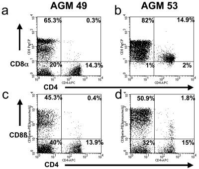
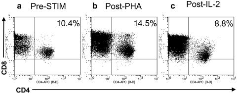
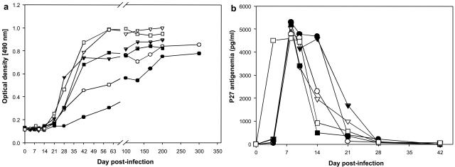
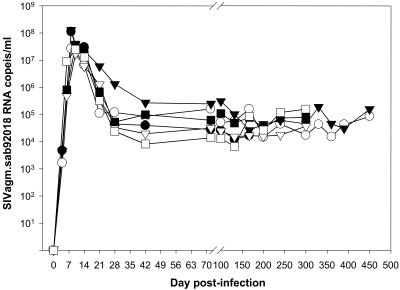
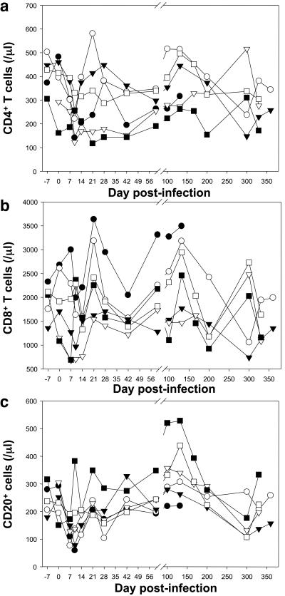
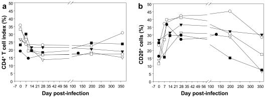
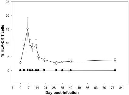
Caribbean-born African green monkeys (AGMs) were classified as Chlorocebus sabaeus by cytochrome b sequencing. Guided by these phylogenetic analyses, we developed a new model for the study of simian immunodeficiency virus (SIV) infection in natural hosts by inoculating Caribbean AGMs with their species-specific SIVagm.sab. SIVagm.sab replicated efficiently in Caribbean AGM peripheral blood mononuclear cells in vitro. During SIVagm.sab primary infection of six Caribbean AGMs, the virus replicated at high levels, with peak viral loads (VLs) of 10(7) to 10(8) copies/ml occurring by day 8 to 10 postinfection (p.i.). Set-point values of up to 2 x 10(5) copies/ml were reached by day 42 p.i. and maintained throughout follow-up (through day 450 p.i.). CD4(+) T-cell counts in the blood showed a transient depletion at the peak of VL, and then returned to near preinfection values by day 28 p.i. and remained relatively stable during the chronic infection. Preservation of CD4 T cells was also found in lymph nodes (LNs) of chronic SIVagm.sab-infected Caribbean AGMs. No activation of CD4(+) T cells was detected in the periphery in SIV-infected Caribbean AGMs. These virological and immunological profiles from peripheral blood and LNs were identical to those previously reported in African-born AGMs infected with the same viral strain (SIVagm.sab92018). Due to these similarities, we conclude that Caribbean AGMs are a useful alternative to AGMs of African origin as a model for the study of SIV infection in natural African hosts.
Figures

Similar articles
-
Experimental depletion of CD8+ cells in acutely SIVagm-infected African Green Monkeys results in increased viral replication.Retrovirology. 2010 May 11;7:42. doi: 10.1186/1742-4690-7-42. Retrovirology. 2010. PMID: 20459829 Free PMC article.
-
Effect of B-cell depletion on viral replication and clinical outcome of simian immunodeficiency virus infection in a natural host.J Virol. 2009 Oct;83(20):10347-57. doi: 10.1128/JVI.00880-09. Epub 2009 Aug 5. J Virol. 2009. PMID: 19656874 Free PMC article.
-
Homeostatic cytokines induce CD4 downregulation in African green monkeys independently of antigen exposure to generate simian immunodeficiency virus-resistant CD8αα T cells.J Virol. 2014 Sep;88(18):10714-24. doi: 10.1128/JVI.01331-14. Epub 2014 Jul 2. J Virol. 2014. PMID: 24991011 Free PMC article.
-
SIVagm: genetic and biological features associated with replication.Front Biosci. 2003 Sep 1;8:d1170-85. doi: 10.2741/1130. Front Biosci. 2003. PMID: 12957815 Review.
-
What can natural infection of African monkeys with simian immunodeficiency virus tell us about the pathogenesis of AIDS?AIDS Rev. 2004 Jan-Mar;6(1):40-53. AIDS Rev. 2004. PMID: 15168740 Review.
Cited by
-
Refinement of a protocol for the induction of lactation in nonpregnant nonhuman primates by using exogenous hormone treatment.J Am Assoc Lab Anim Sci. 2014 Nov;53(6):700-7. J Am Assoc Lab Anim Sci. 2014. PMID: 25650978 Free PMC article.
-
The Hitchhiker Guide to CD4+ T-Cell Depletion in Lentiviral Infection. A Critical Review of the Dynamics of the CD4+ T Cells in SIV and HIV Infection.Front Immunol. 2021 Jul 21;12:695674. doi: 10.3389/fimmu.2021.695674. eCollection 2021. Front Immunol. 2021. PMID: 34367156 Free PMC article.
-
Mucosal simian immunodeficiency virus transmission in African green monkeys: susceptibility to infection is proportional to target cell availability at mucosal sites.J Virol. 2012 Apr;86(8):4158-68. doi: 10.1128/JVI.07141-11. Epub 2012 Feb 8. J Virol. 2012. PMID: 22318138 Free PMC article.
-
Nonprogressive and progressive primate immunodeficiency lentivirus infections.Immunity. 2010 Jun 25;32(6):737-42. doi: 10.1016/j.immuni.2010.06.004. Immunity. 2010. PMID: 20620940 Free PMC article. Review.
-
Lack of Specific Regulatory T Cell Depletion and Cytoreduction Associated with Extensive Toxicity After Administration of Low and High Doses of Cyclophosphamide.AIDS Res Hum Retroviruses. 2022 Jan;38(1):45-49. doi: 10.1089/AID.2021.0036. Epub 2021 Jun 8. AIDS Res Hum Retroviruses. 2022. PMID: 33957772 Free PMC article.
References
-
- Anderson, S., A. T. Bankier, B. G. Barrell, M. H. de Bruijn, A. R. Coulson, J. Drouin, I. C. Eperon, D. P. Nierlich, B. A. Roe, F. Sanger, P. H. Schreier, A. J. Smith, R. Staden, and I. G. Young. 1981. Sequence and organization of the human mitochondrial genome. Nature 290:457-465. - PubMed
-
- Apetrei, C., B. Gormus, I. Pandrea, M. Metzger, P. ten Haaft, L. N. Martin, R. Bohm, X. Alvarez, G. Koopman, M. Murphey-Corb, R. S. Veazey, A. A. Lackner, G. Baskin, J. Heeney, and P. A. Marx. 2004. Direct inoculation of simian immunodeficiency virus from sooty mangabeys in black mangabeys (Lophocebus aterrimus): first evidence of AIDS in a heterologous African species and different pathologic outcomes of experimental infection. J. Virol. 78:11506-11518. - PMC - PubMed
-
- Beer, B., J. Denner, C. R. Brown, S. Norley, J. zur Megede, C. Coulibaly, R. Plesker, S. Holzammer, M. Baier, V. M. Hirsch, and R. Kurth. 1998. Simian immunodeficiency virus of African green monkeys is apathogenic in the newborn natural host. J. Acquir. Immune Defic. Syndr. Hum. Retrovirol. 18:210-220. - PubMed
MeSH terms
Substances
Grants and funding
LinkOut - more resources
Full Text Sources
Research Materials
