. 2006 May;80(10):4927-39.
doi: 10.1128/JVI.80.10.4927-4939.2006.

Adeno-associated virus type 2 increases proteosome-dependent degradation of p21WAF1 in a human papillomavirus type 31b-positive cervical carcinoma line

Affiliations

Adeno-associated virus type 2 increases proteosome-dependent degradation of p21WAF1 in a human papillomavirus type 31b-positive cervical carcinoma line

Samina Alam et al. J Virol. 2006 May.

Abstract

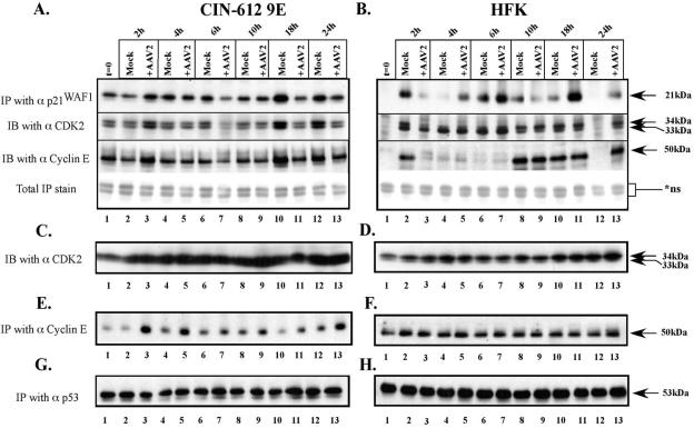
Adeno-associated virus type 2 (AAV2) seropositivity is negatively correlated with the development of human papillomavirus (HPV)-associated cervical cancer. We have begun analysis of the molecular mechanisms underlying AAV2-mediated onco-suppression through cell cycle regulation in HPV-infected keratinocytes isolated from a low-grade cervical lesion. AAV2 superinfection of HPV type 31b (HPV31b)-positive cells at early times postinfection resulted in degradation of the cyclin-dependent kinase (CDK) inhibitor p21(WAF1) protein in a proteosome-dependent manner. Downstream consequences of lowering p21(WAF1) levels included a proportional loss of cyclin E/CDK2 complexes bound to p21(WAF1). The loss of stable p21(WAF1)/cyclin E/CDK2 complexes coincided with an increase in CDK2-associated kinase activity and cyclin E levels. Both events have the potential to enhance the G(1)/S transition point mediated by active cyclin E/CDK2 complexes. Concurrently, cyclin A and E2F levels were decreased, conditions reminiscent of delayed entrance into the S phase of the cell cycle. On the other hand, infection of primary human foreskin keratinocytes with AAV2 resulted in upregulation of p21(WAF1) protein levels, reminiscent of a block in G(1) phase progression. We propose that by down regulating p21(WAF1), AAV2 initiates cell cycle activities leading to enhanced G(1)/S phase-like conditions which may be favorable for AAV2-specific functions and may lead to downstream interference with HPV-associated cervical cancer progression.


Figures

FIG. 1.
FIG. 1.
Cell synchronization. CIN-612 9E (A) and HFK (B) cells were maximally synchronized in the G1 phase of the cell cycle by plating and incubation for 10 to 12 h as described in Materials and Methods. At t = 0, cells were trypsinized and prepared for FACS analysis as described in the text. Confluent CIN-612 9E cells (C) and confluent HFK cells (D) used for the synchronization experiments were prepared for FACS analysis as described in the text.
FIG. 2.
FIG. 2.
AAV2 downregulates p21WAF1 protein levels in HPV-positive keratinocytes. CIN-612 9E (A) and HFK (B) cell lines were infected with AAV2 at an MOI of 100, trypsinized, and pelleted at the stated time points within a 24-h period. Whole-cell extracts were prepared, and 30 μg of total protein sample was resolved on a 12% SDS-polyacrylamide gel, transferred to nitrocellulose membrane, and probed with p21WAF1 polyclonal antibodies as indicated. To detect p27KIP1 protein expression, 30 μg of whole-cell extract was resolved on a 10% SDS-polyacrylamide gel and probed with a polyclonal antibody against p27KIP1 as described for p21WAF1. To detect actin expression, 30 μg of whole-cell extract was resolved on an 8% SDS-polyacrylamide gel and probed with a monoclonal antibody against actin as described for p21WAF1.
FIG. 3.
FIG. 3.
Densitometric analysis of immunoprecipitated p21WAF1 protein levels in AAV2-infected and uninfected CIN-612 9E and HFK cell lines. CIN-612 9E (A) and HFK (B) cell lines were infected with AAV2 as described in Materials and Methods. Cell extracts were prepared, followed by immunoprecipitation with the antibody against p21WAF1. Immunoprecipitates were resolved on a 12% SDS-polyacrylamide gel and transferred to nitrocellulose, and Western blotting was performed with the polyclonal antibody against p21WAF1. Densitometric analysis of bands corresponding to the immunoprecipitated p21WAF1 was performed. Data presented are the means of three experiments. At each time point, immunoprecipitated p21WAF1 protein levels in mock-infected samples were normalized to 1.
FIG. 4.
FIG. 4.
AAV2-mediated downregulation of p21WAF1 levels in HPV-infected cells correlates with the loss of bound CDK2 and cyclin E subunits. (A and B) CIN-612 9E cells (A) and HFK cells (B) were infected with AAV2 and cell extracts were prepared, followed by immunoprecipitation with the p21WAF1 antibody and probing as described in Materials and Methods. To detect bound CDK2 and cyclin subunits, the p21WAF1 immunoprecipitates were examined using Western blotting with polyclonal and monoclonal antibodies against CDK2 and cyclin E, respectively. A 10% and a 7.5% SDS-polyacrylamide gel were used for CDK2 and cyclin E, respectively. To determine equal loading of total protein in immunoprecipitated samples, an identical blot was stained with the GelCode Blue stain reagent. ns, nonspecific. (C and D) CIN-612 9E and HFK cells were infected with AAV2, and whole-cell extracts were prepared as described in Materials and Methods. Total levels of CDK2 were detected using 30 μg of whole-cell extract and resolved on a 10% SDS-polyacrylamide gel, followed by Western blotting using polyclonal antibodies against CDK2. (E and F) CIN-612 9E and HFK cells were infected with AAV2, and cell extracts were prepared as described in the text. Cyclin E protein levels were determined by immunoprecipitating with monoclonal antibody against the cyclin E protein. Immunoprecipitates were resolved on a 7.5% SDS-polyacrylamide gel, and Western blotting was performed to detect cyclin E expression as described in Materials and Methods. (G and H) CIN-612 9E and HFK cells were infected with AAV2, cell extracts were prepared, and immunoprecipitations were performed with antibodies against p53. Immunoprecipitates were resolved on a 7.5% SDS-polyacrylamide gel and transferred to nitrocellulose, and Western blotting was performed using a monoclonal antibody against p53.
FIG. 5.
FIG. 5.
AAV2 targets p21WAF1 for accelerated proteosome-mediated degradation. CIN-612 9E cells were infected with AAV2 as described in Materials and Methods. A duplicate set of cells infected with AAV2 was treated with MG132 proteosome inhibitor at a final concentration of 5 μM. Cell extracts were prepared and used to immunoprecipitate p21WAF1 as described in Materials and Methods. Immunoprecipitates were resolved on a 12% SDS-polyacrylamide gel and transferred to nitrocellulose, followed by Western blotting as discussed above using polyclonal antibodies against p21WAF1.
FIG. 6.
FIG. 6.
CIN-612 9E cells display increased CDK2-associated kinase activity in response to AAV2 infection. CIN-612 9E cells (top panel) and HFK cells (bottom panel) were infected with AAV2 as described in Materials and Methods. Cell extracts were prepared, and immunoprecipitations were performed utilizing a polyclonal antibody against CDK2. Immunoprecipitates were subjected to kinase reactions as described in Materials and Methods, resolved on a 12% polyacrylamide gel, and autoradiographed.
FIG. 7.
FIG. 7.
AAV2 upregulates cyclin E protein levels but downregulates cyclin A and E2F protein levels in HPV-positive cells. CIN-612 9E (A) and HFK (B) cells were infected with AAV2 as described in Materials and Methods. Protein extracts were prepared, followed by immunoprecipitation with monoclonal antibodies against cyclin E and E2F and polyclonal antibodies against cyclin A. Cyclin E and E2F immunoprecipitates were resolved on a 7.5% SDS-polyacrylamide gel, and cyclin A immunoprecipitates were resolved on a 6% SDS-polyacrylamide gel. Reaction mixtures were transferred to nitrocellulose, and Western blotting was performed to detect expression of the respective proteins in response to AAV2. For determining pRb expression, cells were infected as described above and total protein extracts were prepared as described in Materials and Methods. For detecting pRb expression, 60 μg of whole-cell extract was resolved on a 6% SDS-polyacrylamide gel and transferred to nitrocellulose. Western blotting was performed to detect pRb expression using a polyclonal antibody against pRb.
FIG. 8.
FIG. 8.
HPV31:xb cells display decreased p21WAF1 protein levels and increased CDK2-associated kinase activity in response to AAV2 infection. Cutaneous foreskin keratinocyte-derived HPV31:xb cells were infected with AAV2, and cell extracts were prepared for immunoprecipitation as described in Materials and Methods. (A) Immunoprecipitations were performed utilizing a polyclonal antibody against p21WAF1. To determine equal loading of total protein in immunoprecipitated samples, an identical blot was stained with the GelCode Blue stain reagent. ns, nonspecific. (B) Top: immunoprecipitations were performed utilizing a polyclonal antibody against CDK2. Immunoprecipitates were subjected to kinase reactions as described in Materials and Methods, resolved on a 12% polyacrylamide gel, and autoradiographed. Bottom: to detect CDK2 expression, 30 μg of whole-cell extract was resolved on a 10% SDS-polyacrylamide gel and probed with a polyclonal antibody against CDK2.
FIG. 9.
FIG. 9.
Infection of HPV-positive (A) as well as HFK (B) cells with AAV2 does not result in significant changes in cell cycle progression. Both HPV and HFK cells were infected with AAV2 and subjected to FACS analysis as described in Materials and Methods. The percentages of cells in the various cell cycle phases were plotted at the various time points indicated. (C) Complete FACS analysis profiles of AAV2-infected and mock-infected CIN-612 9E cells. (D) Complete FACS analysis profiles of AAV2-infected and mock-infected HFK cells.

Similar articles

Cited by

References

    1. Adams, P. D., and W. G. Kaelin, Jr. 1996. The cellular effects of E2F overexpression. Curr. Top. Microbiol. Immunol. 208:79-93. - PubMed
    1. Bantel-Schaal, U. 1995. Growth properties of a human melanoma cell line are altered by adeno-associated parvovirus type 2. Int. J. Cancer 60:269-274. - PubMed
    1. Bantel-Schaal, U., and H. zur Hausen. 1984. Characterization of the DNA of a defective human parvovirus isolated from a genital site. Virology 134:52-63. - PubMed
    1. Batchu, R. B., M. A. Shammas, J. Y. Wang, J. Freeman, N. Rosen, and N. C. Munshi. 2002. Adeno-associated virus protects the retinoblastoma family of proteins from adenoviral-induced functional inactivation. Cancer Res. 62:2982-2985. - PubMed
    1. Batchu, R. B., M. A. Shammas, J. Y. Wang, and N. C. Munshi. 2001. Dual level inhibition of E2F-1 activity by adeno-associated virus Rep78. J. Biol. Chem. 276:24315-24322. - PubMed

Publication types

MeSH terms

Substances