Comparative Study
. 2006 May;80(10):4940-8.
doi: 10.1128/JVI.80.10.4940-4948.2006.

Different domains of CD81 mediate distinct stages of hepatitis C virus pseudoparticle entry

Affiliations
Comparative Study

Different domains of CD81 mediate distinct stages of hepatitis C virus pseudoparticle entry

Claire Bertaux et al. J Virol. 2006 May.

Abstract

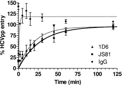
The CD81 tetraspanin was first identified as a hepatitis C virus (HCV) receptor by its ability to bind the soluble ectodomain of envelope glycoprotein E2 (sE2). More recently, it has been suggested that CD81 is necessary but not sufficient for HCV entry into target cells. Here we present further evidence that putative human hepatocyte-specific factors act in concert with CD81 to mediate sE2 binding and HCV pseudoparticle (HCVpp) entry. Moreover, we show that CD81-mediated HCVpp entry entails E2 binding to residues in the large extracellular loop as well as molecular events mediated by the transmembrane and intracellular domains of CD81. The concept that CD81 receptor function progresses in stages is further supported by our finding that anti-CD81 monoclonal antibodies inhibit HCVpp entry by different mechanisms. The half-life of CD81-HCVpp binding was determined to be approximately 17 min, and we propose that binding is followed by CD81 oligomerization, partitioning into cholesterol-rich membrane domains, or other, lateral protein-protein interactions. This results in the formation of a receptor-virus complex that undergoes endocytosis and pH-dependent membrane fusion.


Figures

FIG. 1.
FIG. 1.
Expression of CD81 on the cell surface and sE2 binding. (A) CD81 expression was determined with different cell lines, including human hepatoma cells permissive to HCVpp (checkered bars), human cells nonpermissive to HCVpp (solid bars), and murine cells (hatched bars), as indicated along the x axis. CD81 was quantified by flow cytometry (MFI) after labeling with an anti-CD81 MAb (JS81). Results are means of three independent experiments ± standard deviations (SD). Permissivity (+, 104 to 105 RLU; ++, 105 to 106 RLU) or resistance (−) of cells to HCVpp is indicated under the bar graph. (B) sE2 binding to cells from panel A, as indicated along the x axis, was quantified by flow cytometry (MFI) after labeling of cells with the anti-E2 MAb H53 followed by a PE-conjugated anti-mouse IgG antibody. Results are means of three independent experiments ± SD. (C) Anti-CD81 MAbs JS81 (black bars) and 1D6 (hatched bars) were used to inhibit sE2 binding to a subset of cells from panel A. Residual sE2 on cells was quantified by flow cytometry (MFI) after labeling with H53-coupled fluorescent beads. The percentage of inhibition of sE2 binding is indicated for each cell line and was calculated relative to sE2 binding in the presence of a nonspecific isotype-matched murine IgG. Values are means of three independent experiments ± standard deviations.
FIG. 1.
FIG. 1.
Expression of CD81 on the cell surface and sE2 binding. (A) CD81 expression was determined with different cell lines, including human hepatoma cells permissive to HCVpp (checkered bars), human cells nonpermissive to HCVpp (solid bars), and murine cells (hatched bars), as indicated along the x axis. CD81 was quantified by flow cytometry (MFI) after labeling with an anti-CD81 MAb (JS81). Results are means of three independent experiments ± standard deviations (SD). Permissivity (+, 104 to 105 RLU; ++, 105 to 106 RLU) or resistance (−) of cells to HCVpp is indicated under the bar graph. (B) sE2 binding to cells from panel A, as indicated along the x axis, was quantified by flow cytometry (MFI) after labeling of cells with the anti-E2 MAb H53 followed by a PE-conjugated anti-mouse IgG antibody. Results are means of three independent experiments ± SD. (C) Anti-CD81 MAbs JS81 (black bars) and 1D6 (hatched bars) were used to inhibit sE2 binding to a subset of cells from panel A. Residual sE2 on cells was quantified by flow cytometry (MFI) after labeling with H53-coupled fluorescent beads. The percentage of inhibition of sE2 binding is indicated for each cell line and was calculated relative to sE2 binding in the presence of a nonspecific isotype-matched murine IgG. Values are means of three independent experiments ± standard deviations.
FIG. 2.
FIG. 2.
CD81 sequence and structure. (A) Comparison of human, mouse, and rat CD81 LEL sequences. Residues differing between human and mouse CD81 proteins are shaded in gray. Most of these residues are also different in rat CD81 and are therefore not indicated. The two additional residues that differ in rat CD81 are shaded in black. (B) Predicted two-dimensional structure of CD81, including the two extracellular loops, four transmembrane helices, and the intracellular loop along with the intracellular N-terminal and C-terminal tails. LEL residues that were mutagenized in this study are shown using the same color code as that in panel A. Polar transmembrane residues and intracellular cysteines that were mutagenized are indicated in white circles and squares, respectively.
FIG. 3.
FIG. 3.
Role of specific CD81 LEL residues in HCVpp entry and sE2 binding. (A) HCVpp entry was tested in HepG2 cells transfected with pcDNA3.1, human wild-type CD81, murine wild-type CD81 (all in white bars), and human CD81 mutants, as indicated along the x axis. Human residues were replaced by alanine (black bars) or by corresponding murine (shaded bars) or rat (hatched bars) CD81 residues. HCVpp entry was calculated as a percentage of the entry level into cells expressing human wild-type CD81. The expression of CD81 mutants was quantified by flow cytometry after labeling of cells with JS81 (or 1D6 for the A164T mutant) and was expressed as a percentage of human wild-type CD81 expression. The percentage of HCVpp entry was further normalized for CD81 expression. Values are means of three independent experiments ± SD. (B) Binding of sE2 to CD81 mutants with significantly decreased HCVpp entry was quantified by flow cytometry. HepG2 cells were transfected with pcDNA3.1, human wild-type CD81, murine CD81, and CD81 mutants. Transfected cells were incubated with sE2, followed by labeling with H53 and a PE-conjugated anti-mouse IgG. Binding was analyzed by flow cytometry and expressed as a percentage of sE2 binding to HepG2 cells expressing human wild-type CD81. The percentage of sE2 binding was further normalized for mutant expression levels. Values are averages of three independent experiments ± SD.
FIG. 4.
FIG. 4.
Role of specific CD81 transmembrane and intracytoplasmic residues in HCVpp entry and sE2 binding. (A) HCVpp entry was tested in HepG2 cells transfected with pcDNA3.1 (white bars), human wild-type CD81 (white bars), or CD81 mutants (black bars), as indicated along the x axis. HCVpp entry was calculated as a percentage of the entry level into cells expressing human wild-type CD81. The expression of CD81 mutants was quantified by flow cytometry after labeling of cells with JS81 and was expressed as a percentage of human wild-type CD81 expression. The percentage of HCVpp entry was further normalized for CD81 expression. Values are means of three independent experiments ± SD. (B) Binding of sE2 to CD81 mutants with significantly decreased HCVpp entry was quantified by flow cytometry. HepG2 cells were transfected with pcDNA3.1, human wild-type CD81, and CD81 mutants. Transfected cells were incubated with sE2, followed by labeling with H53 and PE-conjugated anti-mouse IgG. Binding was analyzed by flow cytometry and expressed as a percentage of E2 binding to HepG2 cells expressing human wild-type CD81. The percentage of sE2 binding was further normalized for mutant expression levels. Values are means of three independent experiments ± SD.
FIG. 5.
FIG. 5.
Binding to cells and inhibition of HCVpp entry by anti-CD81 MAbs. (A) Different concentrations of MAbs JS81 (squares) and 1D6 (triangles) ranging over 2 orders of magnitude were added to Huh-7 cells, and binding was quantified by flow cytometry (MFI) after cell labeling with a PE-conjugated anti-mouse IgG. Binding is expressed as a percentage of the MFI at the highest, saturating concentration of MAb, beyond which no further effect was observed. Values are means of three independent experiments ± SD. (B) Different concentrations of MAbs JS81 (squares) and 1D6 (triangles) were added to Huh-7 cells, which were then infected with HCVpp. Luciferase activity was measured in cell lysates 48 h after infection. The percentage of HCVpp entry was calculated relative to entry in the absence of anti-CD81 MAbs. A 100% inhibition of HCVpp entry was set at the MAb concentration beyond which no further effect was observed. Values are means of three independent experiments ± SD. (C) Calculated EC50 and IC50 values for JS81 and 1D6 after nonlinear curve fitting using GraphPad Prism 4 software.
FIG. 6.
FIG. 6.
Time course of inhibition of HCVpp entry. Anti-CD81 MAbs were added to cells that were prebound to viral particles by centrifugation at 4°C. Luciferase activities were measured at 48 h postinfection and expressed relative to entry in the absence of antibody. Values are means of three independent experiments ± SD.

References

    1. Allander, T., X. Forns, S. U. Emerson, R. H. Purcell, and J. Bukh. 2000. Hepatitis C virus envelope protein E2 binds to CD81 of tamarins. Virology 277:358-367. - PubMed
    1. Bartosch, B., J. Dubuisson, and F. L. Cosset. 2003. Infectious hepatitis C virus pseudo-particles containing functional E1-E2 envelope protein complexes. J. Exp. Med. 197:633-642. - PMC - PubMed
    1. Bartosch, B., A. Vitelli, C. Granier, C. Goujon, J. Dubuisson, S. Pascale, E. Scarselli, R. Cortese, A. Nicosia, and F. L. Cosset. 2003. Cell entry of hepatitis C virus requires a set of co-receptors that include the CD81 tetraspanin and the SR-B1 scavenger receptor. J. Biol. Chem. 278:41624-41630. - PubMed
    1. Berditchevski, F. 2001. Complexes of tetraspanins with integrins: more than meets the eye. J. Cell Sci. 114:4143-4151. - PubMed
    1. Berditchevski, F., K. F. Tolias, K. Wong, C. L. Carpenter, and M. E. Hemler. 1997. A novel link between integrins, transmembrane-4 superfamily proteins (CD63 and CD81), and phosphatidylinositol 4-kinase. J. Biol. Chem. 272:2595-2598. - PubMed

Publication types

MeSH terms

LinkOut - more resources