. 2006 May;80(10):5010-20.
doi: 10.1128/JVI.80.10.5010-5020.2006.

Cellular splicing and transcription regulatory protein p32 represses adenovirus major late transcription and causes hyperphosphorylation of RNA polymerase II

Affiliations

Cellular splicing and transcription regulatory protein p32 represses adenovirus major late transcription and causes hyperphosphorylation of RNA polymerase II

Christina Ohrmalm et al. J Virol. 2006 May.

Abstract

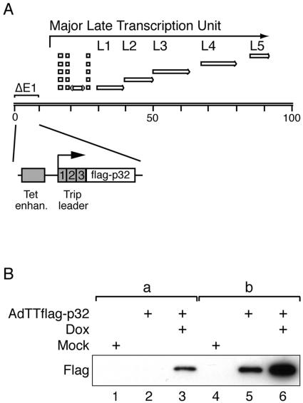
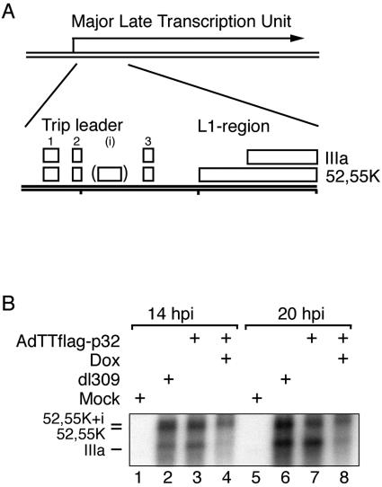
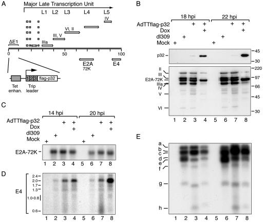
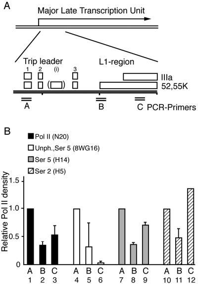
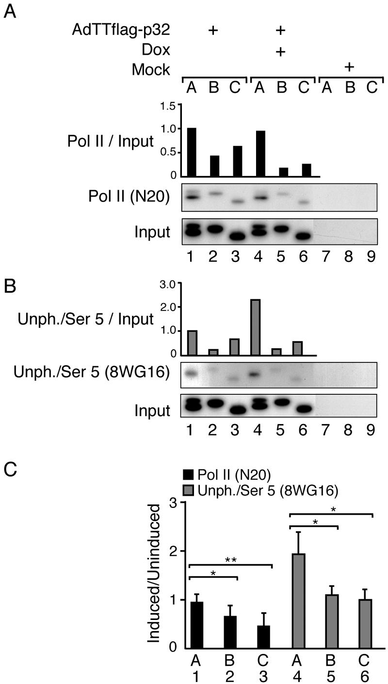
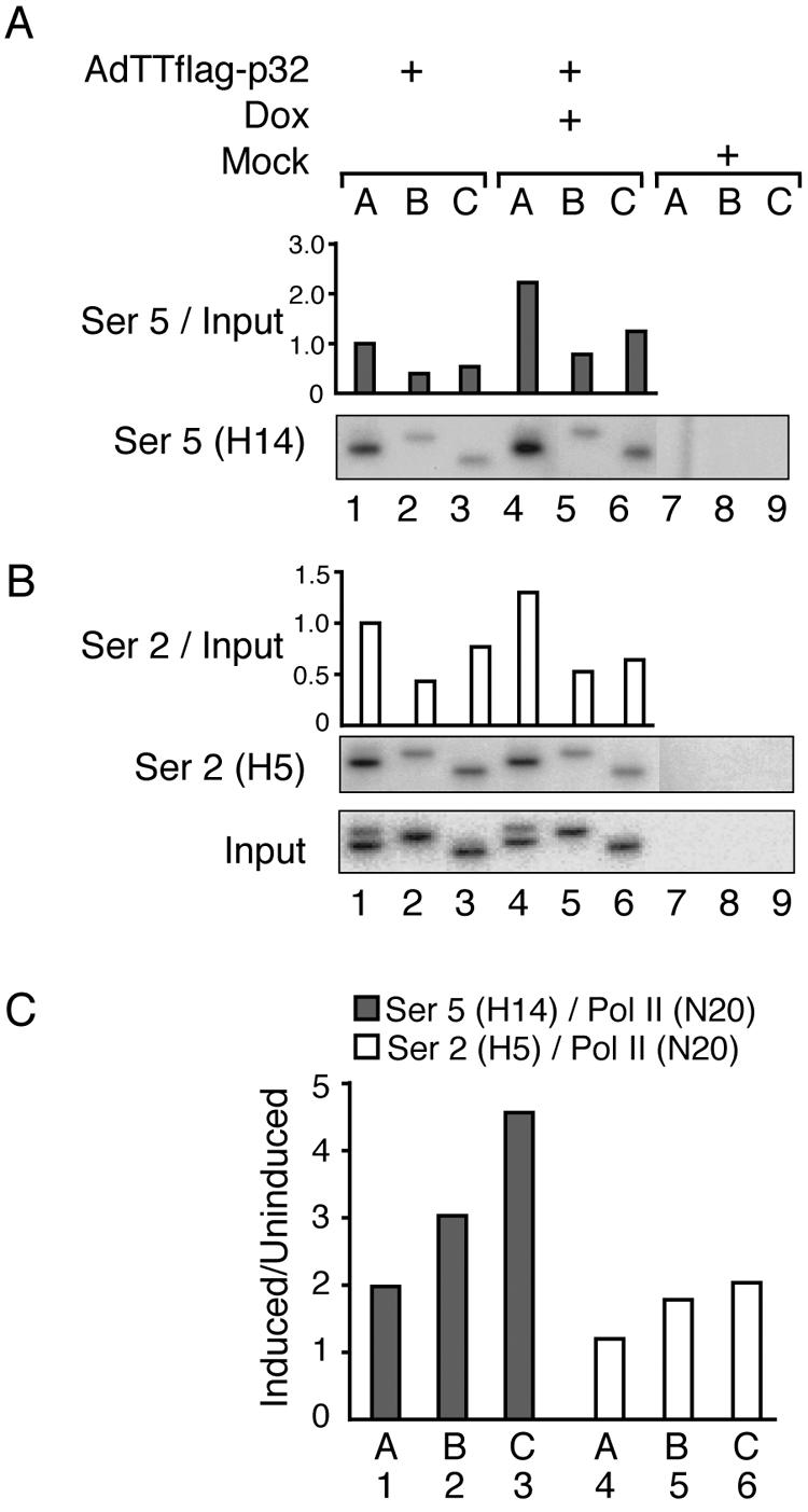
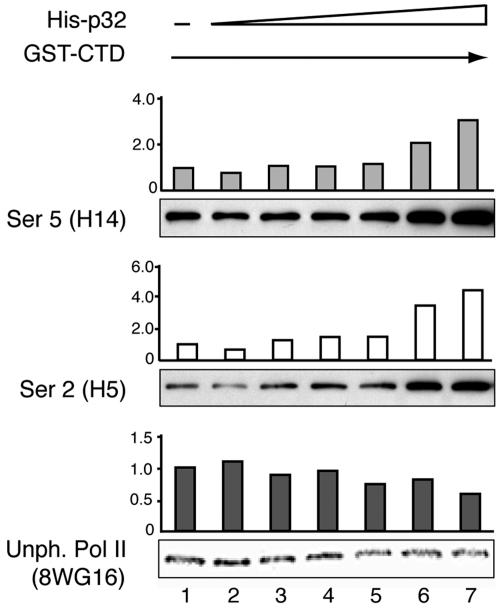
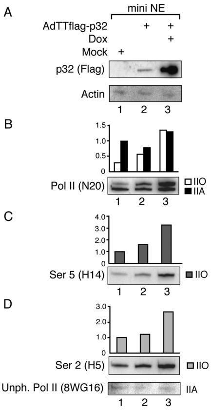
The cellular protein p32 is a multifunctional protein, which has been shown to interact with a large number of cellular and viral proteins and to regulate several important activities like transcription and RNA splicing. We have previously shown that p32 regulates RNA splicing by binding and inhibiting the essential SR protein ASF/SF2. To determine whether p32 also functions as a regulator of splicing in virus-infected cells, we constructed a recombinant adenovirus expressing p32 under the transcriptional control of an inducible promoter. Much to our surprise the results showed that p32 overexpression effectively blocked mRNA and protein expression from the adenovirus major late transcription unit (MLTU). Interestingly, the p32-mediated inhibition of MLTU transcription was accompanied by an approximately 4.5-fold increase in Ser 5 phosphorylation and an approximately 2-fold increase in Ser 2 phosphorylation of the carboxy-terminal domain (CTD). Further, in p32-overexpressing cells the efficiency of RNA polymerase elongation was reduced approximately twofold, resulting in a decrease in the number of polymerase molecules that reached the end of the major late L1 transcription unit. We further show that p32 stimulates CTD phosphorylation in vitro. The inhibitory effect of p32 on MLTU transcription appears to require the CAAT box element in the major late promoter, suggesting that p32 may become tethered to the MLTU via an interaction with the CAAT box binding transcription factor.


Figures

FIG. 1.
FIG. 1.
Induction of Flag-p32 expression. (A) Schematic illustration showing the structure of recombinant virus AdTTflag-p32. The tetracycline-regulatable gene cassette is located at the left end of the viral genome, replacing all of the E1A region and most of the E1B region. The nucleotide sequence of this construct is available upon request. The structure of the coterminal mRNA families derived from the major late transcription unit is also indicated. (B) Western blot analysis of Flag-p32 expression in AdTTflag-p32-infected 293 cells harvested 20 hpi. In groups a (lanes 1 to 3) and b (lanes 4 to 6), 0.4 μg and 1.6 μg of extract, respectively, was separated by SDS-PAGE and probed with an anti-Flag monoclonal antibody. Note that cells infected with the AdTTflag-p32 virus were also coinfected with an equal amount (5 FFU per cell) of the activator virus AdCMVrtTA. The coinfection protocol is necessary in order to provide the reverse tetracycline activator protein that drives Flag-p32 expression (34).
FIG. 2.
FIG. 2.
p32 overexpression blocks L1 mRNA accumulation. (A) Schematic illustration showing the spliced structure of the 52,55K and the IIIa mRNAs expressed from region L1. (B) Northern blot analysis of total cytoplasmic RNA prepared from AdTTflag-p32-infected cells at the indicated time points. RNA was transferred to a nitrocellulose filter and hybridized with a 32P-labeled probe specific for region L1. Note that cells infected with the AdTTflag-p32 virus were also coinfected with an equal amount (5 FFU per cell) of the activator virus AdCMVrtTA. The coinfection protocol is necessary in order to provide the reverse tetracycline activator protein that drives Flag-p32 expression (34).
FIG. 3.
FIG. 3.
p32 overexpression blocks gene expression from the MLTU. (A) Schematic illustration showing the position of MLTU units L1 to L5, regions E2A and E4, with the positions of the coding regions for proteins relevant for this study indicated. AdTTflag-p32-infected 293 cells were harvested at the time points indicated, and protein (B), total cytoplasmic RNA (C and D), and viral DNA (E) were isolated and analyzed as described in Materials and Methods. (B) Western blot analysis using an anti-Flag monoclonal antibody (upper part) or an antibody directed against late viral proteins (lower part). The positions of the Flag-p32 protein, late structural proteins, and the E2A-72K DNA binding protein are indicated on the left. (C) Northern blot analysis using a 32P-labeled probe specific for region E2A. (D) Northern blot analysis using a 32P-labeled probe specific for region E4. Sizes of the RNA classes are given in kilobases (47). (E) Southern blot analysis of HindIII-digested low-molecular-weight DNA (fragments a to h) using a 32P-labeled probe prepared by random priming of dl309 virion DNA.
FIG. 4.
FIG. 4.
p32 inhibition of major late promoter activity requires the CAAT box. (A) Increasing amounts of pCMV-p32 (0.05, 0.1, 0.5, or 1 μg) as well as an empty CMV control vector (−) were cotransfected together with a constant amount (1 μg) of the wild-type reporter construct pTrip-CAT (bars 6 to 10), CAAT box mutant pTripCAT (CCCAT) (bars 11 to 15), or no CAT-expressing plasmid (bars 1 to 5). Cell extracts were prepared between 24 and 36 h posttransfection, and CAT protein expression was quantitated by CAT ELISA (Boehringer Mannheim). (B) Schematic illustration showing the structures of the tripeptidyl-peptidase II promoter luciferase constructs (23). The full-length construct (174) contains the two inverted CAAT boxes and the initiator (Inr) element found in the wild-type promoter (23), whereas constructs 104 and 130 contained only one of the CAAT boxes. Construct 118 lacks both CAAT boxes, and the empty pGL3-basic vector was used as a negative control (−). (C) A constant amount of the different luciferase constructs (1 μg) was cotransfected with an empty CMV control vector (0.2 μg; black bars) or with pCMV-p32 (0.2 μg; gray bars). Cell extracts were prepared at 48 h posttransfection, and luciferase protein expression was quantified in a Luminoscan RT (Labsystems). The results represent means ± standard deviations from three independent transfections.
FIG. 5.
FIG. 5.
The cyclic phosphorylation of the CTD during a wild-type-adenovirus infection. (A) Schematic figure showing an expansion of the L1 transcription unit with the positions of the spliced tripartite leader segments and the L1 52,55K and IIIa mRNA bodies indicated (for a review see reference 15). The approximate positions of the three PCR primer pairs used in the ChIP assay are indicated. (B) Densities of different phosphorylated forms of RNA pol II along the MLTU L1 region. A panel of four antibodies recognizing either the total number of RNA pol II or different forms of phosphorylated CTD were used in the ChIP assay. The results from the PCR amplifications were quantitated by PhosphorImager analysis and are presented here in relation to the value obtained from PCR primer pair A (promoter proximal), which is set as 1 for each antibody.
FIG. 6.
FIG. 6.
p32 overexpression reduces the efficiency of RNA Pol II elongation. AdTTflag-p32-infected 293 cells were fixed 22 hpi, and ChIP assays were performed using antibody N20, which recognizes the amino terminus of the large subunit of RNA Pol II (A), or antibody 8WG16, which detects primarily unphosphorylated CTD and weakly Ser 5-phosphorylated CTD (B). Signals were quantitated by PhosphorImager analysis and normalized against the input DNA, which shows the signal from the chromatin before immunoprecipitation. The signal for the promoter (lane 1) in uninduced cells was set as 1 in the respective panels. The data in panels A and B display one representative experiment each. (C) Ratios of the RNA Pol II density at positions A, B, and C (Fig. 5A) in response to p32 induction. Values represent means ± standard deviations from six (N20) or four (8WG16) independent experiments, with a statistically significant reduction (*, P < 0.05; **, P < 0.01) between indicated comparisons (one-tailed paired t test analysis). Note that cells infected with the AdTTflag-p32 virus were also coinfected with an equal amount (5 FFU per cell) of the activator virus AdCMVrtTA. The coinfection protocol is necessary in order to provide the reverse tetracycline activator protein that drives Flag-p32 expression (34).
FIG. 7.
FIG. 7.
p32 overexpression induces CTD hyperphosphorylation. AdTTflag-p32-infected 293 cells were fixed 22 hpi, and ChIP assays were performed using antibody H14, which recognizes Ser 5 phosphorylation (A) or antibody H5, which detects Ser 2-phosphorylated CTD (B). Signals were quantitated by PhosphorImager analysis and normalized against the input DNA, which shows the signal from the chromatin before immunoprecipitation. The signal for the promoter (lane 1) in uninduced cells was set as 1 in the respective panels. The data in panels A and B display the results from one representative experiment. (C) Mean ratio of RNA Pol II density, using Ser 5- or Ser 2-specific antibodies, from several experiments in response to p32 induction divided by the total density of RNA Pol II detected at the respective chromosomal position (for the N20 antibody, data were taken from Fig. 6C). Note that cells infected with the AdTTflag-p32 virus were also coinfected with an equal amount (5 FFU per cell) of the activator virus AdCMVrtTA. The coinfection protocol is necessary in order to provide the reverse tetracycline activator protein that drives Flag-p32 expression (34).
FIG. 8.
FIG. 8.
p32 stimulates CTD phosphorylation in vitro. Increasing amounts of a bacterially expressed His-p32 protein (0, 30, 100, 300, 600, 1,200, and 2,400 pmol) were preincubated in HeLa nuclear extracts and then further incubated with a constant amount of GST-CTD (10 pmol). Proteins were separated on an SDS-PAGE gel, and the extent of CTD phosphorylation was quantitated by PhosphorImager analysis of Western blots probed with phosphoepitope-specific antibodies.
FIG. 9.
FIG. 9.
Overexpression of Flag-p32 increases RNA Pol IIO hyperphosphorylation. Mini-nuclear extracts were prepared from AdTTflag-p32-infected 293 cells 22 hpi or control cells and subjected to Western blot analysis using a panel of antibodies. Note that cells infected with the AdTTflag-p32 virus were also coinfected with an equal amount (5 FFU per cell) of the activator virus AdCMVrtTA. The coinfection protocol is necessary in order to provide the reverse tetracycline activator protein that drives Flag-p32 expression (34).

Similar articles

Cited by

References

    1. Abbott, K. L., J. Archambault, H. Xiao, B. D. Nguyen, R. G. Roeder, J. Greenblatt, J. G. Omichinski, and P. Legault. 2005. Interactions of the HIV-1 Tat and RAP74 proteins with the RNA polymerase II CTD phosphatase FCP1. Biochemistry 44:2716-2731. - PubMed
    1. Ahn, S. H., M. Kim, and S. Buratowski. 2004. Phosphorylation of serine 2 within the RNA polymerase II C-terminal domain couples transcription and 3′ end processing. Mol. Cell 13:67-76. - PubMed
    1. Ausubel, F. M., R. Brent, R. E. Kingston, D. D. Moore, J. G. Seidman, J. A. Smith, and K. Struhl (ed.). 1995. Current protocols in molecular biology. John Wiley & Sons, Inc., New York, N.Y.
    1. Berenjian, S., and G. Akusjärvi. 2006. Binary AdEasy vector systems designed for Tet-ON or Tet-OFF regulated control of transgene expression. Virus Res. 115:116-123. - PubMed
    1. Chambers, R. S., B. Q. Wang, Z. F. Burton, and M. E. Dahmus. 1995. The activity of COOH-terminal domain phosphatase is regulated by a docking site on RNA polymerase II and by the general transcription factors IIF and IIB. J. Biol. Chem. 270:14962-14969. - PubMed

Publication types

MeSH terms