. 2006 May;80(10):5021-31.
doi: 10.1128/JVI.80.10.5021-5031.2006.

Human rhinovirus attenuates the type I interferon response by disrupting activation of interferon regulatory factor 3

Affiliations

Human rhinovirus attenuates the type I interferon response by disrupting activation of interferon regulatory factor 3

Tao Peng et al. J Virol. 2006 May.

Erratum in

  • J Virol. 2006 Jul;80(13):6722

Retraction in

Abstract

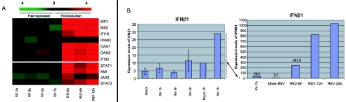
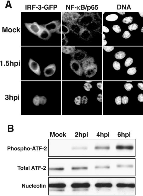
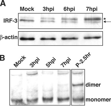
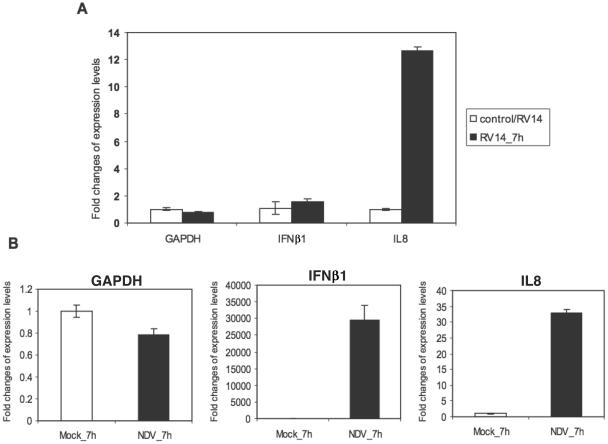
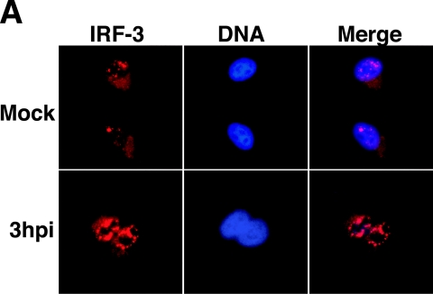
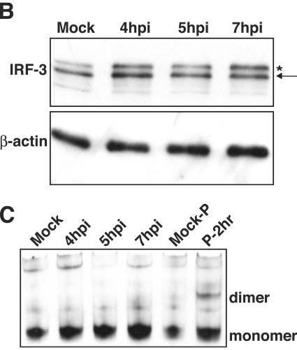
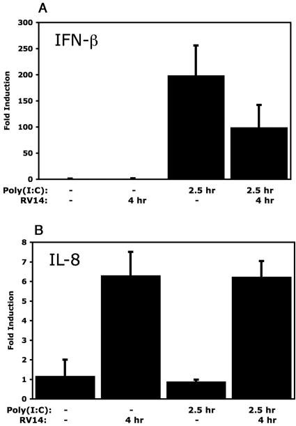
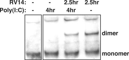
The type I interferon (IFN) response requires the coordinated activation of the latent transcription factors NF-kappaB, interferon regulatory factor 3 (IRF-3), and ATF-2, which in turn activate transcription from the IFN-beta promoter. Synthesis and subsequent secretion of IFN-beta activate the Jak/STAT signaling pathway, resulting in the transcriptional induction of the full spectrum of antiviral gene products. We utilized high-density microarrays to examine the transcriptional response to rhinovirus type 14 (RV14) infection in HeLa cells, with particular emphasis on the type I interferon response and production of IFN-beta. We found that, although RV14 infection results in altered levels of a wide variety of host mRNAs, induction of IFN-beta mRNA or activation of the Jak/STAT pathway is not seen. Prior work has shown, and our results have confirmed, that NF-kappaB and ATF-2 are activated following infection. Since many viruses are known to target IRF-3 to inhibit the induction of IFN-beta mRNA, we analyzed the status of IRF-3 in infected cells. IRF-3 was translocated to the nucleus and phosphorylated in RV14-infected cells. Despite this apparent activation, very little homodimerization of IRF-3 was evident following infection. Similar results in A549 lung alveolar epithelial cells demonstrated the biological relevance of these findings to RV14 pathogenesis. In addition, prior infection of cells with RV14 prevented the induction of IFN-beta mRNA following treatment with double-stranded RNA, indicating that RV14 encodes an activity that specifically inhibits this innate host defense pathway. Collectively, these results indicate that RV14 infection inhibits the host type I interferon response by interfering with IRF-3 activation.


Figures

FIG. 1.
FIG. 1.
RV14 infection of HeLa cells fails to induce a type I interferon response. (A) Comparison of microarray data for various ISG mRNAs from cells infected with RV14 for different periods of time (1, 3, 5, or 7 h postinfection), treated with IFN-α for 6 h (IFN-6h) (8), or infected with RSV for 6 h (RSV-6h) or 12 h (RSV-12h) (62). (B) Raw intensities for IFN-β mRNA, as calculated using MAS5 analysis software, from cells infected with RV14 for 1, 3, 5, or 7 h or with RSV for 6, 12, or 24 h (62). Note that the 7-h time point for RV14-infected cells is shown in both graphs to illustrate the different levels of induction in RV14- and RSV-infected cells.
FIG. 2.
FIG. 2.
Activation of NF-κB, IRF-3, and ATF-2 in RV14-infected cells. (A) Nuclear accumulation of IRF-3 and NF-κB. HeLa cells were transiently transfected with an IRF-3-GFP expression vector and were either mock infected or infected with RV14 and analyzed at 1.5 or 3 hpi. The panels labeled IRF-3-GFP show the localization of the IRF-3-GFP fusion protein, using a fluorescein isothiocyanate filter. The NF-κB/p65 panels show the same field stained with antibodies to detect the p65subunit of NF-κB, visualized using a tetramethyl rhodamine isothiocyanate filter. The DNA panels show the same field stained with Hoechst to reveal cell nuclei. (B) Phosphorylation of ATF-2. Whole-cell lysates (25 μg) prepared from mock-infected cells or cells that had been infected for the indicated amounts of time were analyzed by immunoblotting. ATF-2 was detected by sequentially probing the membrane, using antibodies that recognize the phosphorylated form of ATF-2 (phospho-ATF-2) and all forms of ATF-2 (Total ATF-2). The membrane was also probed with an antibody to nucleolin to show equivalent loading of protein lysates.
FIG. 3.
FIG. 3.
Phosphorylation and homodimerization of IRF-3 in HeLa cells. (A) Phosphorylation of IRF-3. Whole-cell lysates prepared from mock-infected cells or cells that had been infected with RV14 for the indicated amounts of time were analyzed by immunoblotting. IRF-3 was detected by probing the membrane with rabbit polyclonal antibody that detects IRF-3. The nonphosphorylated form of IRF-3 is indicated with an arrow, and the phosphorylated form is indicated with an asterisk. The membrane was stripped and reprobed with an antibody to β-actin to show equivalent loading of protein. (B) Homodimerization of IRF-3. Cell lysates prepared from mock-infected cells or cells that had been infected with RV14 or treated with poly(I:C) for the indicated amounts of time were analyzed by native PAGE, followed by immunoblotting to detect IRF-3. The monomeric and dimeric forms of IRF-3 are indicated.
FIG. 4.
FIG. 4.
Expression of IFN-β mRNA and IL-8 mRNA in RV14-infected A549 cells. (A) Total RNAs extracted from mock-infected or RV14-infected cells were analyzed by qRT-PCR. qRT-PCR data for IFN-β (IFNβ1) and IL-8 (IL8) mRNA are shown, with error bars indicating one standard deviation from the results from three replicates. Results for GAPDH are shown as a normalization control. (B) Total RNAs extracted from mock-infected or NDV-infected A549 cells for the indicated time were analyzed by qRT-PCR. IFN-β, IL-8, and GAPDH mRNAs were examined as described for panel A.
FIG. 5.
FIG. 5.
Analysis of the status of IRF-3 in RV14-infected A549 cells. (A) Subcellular localization of IRF-3. A549 cells were either mock infected or infected with RV14 for the indicated amount of time and were analyzed by immunofluorescence assay. The IRF-3 panels show cells stained with a rabbit polyclonal antibody to detect IRF-3, using a tetramethyl rhodamine isothiocyanate filter. The DNA panels show the same field stained with Hoechst to reveal nuclei. The merged panels show overlays of the IRF-3 and DNA images. (B) Phosphorylation of IRF-3. Whole-cell lysates prepared from mock-infected cells or cells that had been infected with RV14 for the indicated amounts of time were analyzed by immunoblotting. IRF-3 was detected by probing the membrane with rabbit polyclonal antibody that detects human IRF-3. The nonphosphorylated form of IRF-3 is indicated with an arrow, and the phosphorylated form is indicated with an asterisk. The membrane was stripped and reprobed with an antibody to β-actin to show equivalent loading of protein lysates. (C) Homodimerization of IRF-3. Cell lysates prepared from mock-infected cells or cells that had been infected with RV14 or treated with poly(I:C) for the indicated amounts of time were analyzed by native PAGE, followed by immunoblotting to detect IRF-3. The monomeric and dimeric forms of IRF-3 are indicated.
FIG. 5.
FIG. 5.
Analysis of the status of IRF-3 in RV14-infected A549 cells. (A) Subcellular localization of IRF-3. A549 cells were either mock infected or infected with RV14 for the indicated amount of time and were analyzed by immunofluorescence assay. The IRF-3 panels show cells stained with a rabbit polyclonal antibody to detect IRF-3, using a tetramethyl rhodamine isothiocyanate filter. The DNA panels show the same field stained with Hoechst to reveal nuclei. The merged panels show overlays of the IRF-3 and DNA images. (B) Phosphorylation of IRF-3. Whole-cell lysates prepared from mock-infected cells or cells that had been infected with RV14 for the indicated amounts of time were analyzed by immunoblotting. IRF-3 was detected by probing the membrane with rabbit polyclonal antibody that detects human IRF-3. The nonphosphorylated form of IRF-3 is indicated with an arrow, and the phosphorylated form is indicated with an asterisk. The membrane was stripped and reprobed with an antibody to β-actin to show equivalent loading of protein lysates. (C) Homodimerization of IRF-3. Cell lysates prepared from mock-infected cells or cells that had been infected with RV14 or treated with poly(I:C) for the indicated amounts of time were analyzed by native PAGE, followed by immunoblotting to detect IRF-3. The monomeric and dimeric forms of IRF-3 are indicated.
FIG. 6.
FIG. 6.
Inhibition of the dsRNA response in HeLa cells. (A) IFN-β mRNA levels. HeLa cells were either mock infected or infected with RV14 and then treated with poly(I:C) or not for the indicated amounts of time. Total RNA was isolated and analyzed by qRT-PCR for IFN-β mRNA and normalized to levels of rRNA. Error bars indicate one standard deviation from the results from three biological replicates. (B) The RNAs described above were analyzed by qRT-PCR to determine IL-8 mRNA levels.
FIG. 7.
FIG. 7.
Inhibition of the dsRNA response in A549 cells. (A) IFN-β mRNA levels. A549 cells were either mock infected or infected with RV14 and then treated with poly(I:C) or not for the indicated amounts of time. Total RNA was isolated and analyzed by qRT-PCR for IFN-β mRNA and normalized to levels of rRNA. Error bars indicate one standard deviation from the results from three biological replicates. (B) The RNAs described above were analyzed by qRT-PCR to determine IL-8 mRNA levels.
FIG. 8.
FIG. 8.
IRF-3 homodimerization in cells treated with poly(I:C). Whole-cell lysates were prepared from cells that were either mock infected or infected with RV14 and then treated with poly(I:C) or not for the indicated amounts of time. Lysates were analyzed by native PAGE, followed by immunoblotting to detect IRF-3. The monomeric and dimeric forms of IRF-3 are indicated.

Similar articles

Cited by

References

    1. Abzug, M. J., A. C. Beam, E. A. Gyorkos, and M. J. Levin. 1990. Viral pneumonia in the first month of life. Pediatr. Infect. Dis. J. 9:881-885. - PubMed
    1. Baigent, S. J., G. Zhang, M. D. Fray, H. Flick-Smith, S. Goodbourn, and J. W. McCauley. 2002. Inhibition of beta interferon transcription by noncytopathogenic bovine viral diarrhea virus is through an interferon regulatory factor 3-dependent mechanism. J. Virol. 76:8979-8988. - PMC - PubMed
    1. Barro, M., and J. T. Patton. 2005. Rotavirus nonstructural protein 1 subverts innate immune response by inducing degradation of IFN regulatory factor 3. Proc. Natl. Acad. Sci. USA 102:4114-4119. - PMC - PubMed
    1. Basler, C. F., A. Mikulasova, L. Martinez-Sobrido, J. Paragas, E. Muhlberger, M. Bray, H. D. Klenk, P. Palese, and A. Garcia-Sastre. 2003. The Ebola virus VP35 protein inhibits activation of interferon regulatory factor 3. J. Virol. 77:7945-7956. - PMC - PubMed
    1. Bertino, J. S. 2002. Cost burden of viral respiratory infections: issues for formulary decision makers. Am. J. Med. 112(Suppl. 6A):42S-49S. - PubMed

Publication types

MeSH terms