. 2006 Apr 26;34(7):2117-27.
doi: 10.1093/nar/gkl182. Print 2006.

Predictive modelling of topology and loop variations in dimeric DNA quadruplex structures

Affiliations

Predictive modelling of topology and loop variations in dimeric DNA quadruplex structures

Pascale Hazel et al. Nucleic Acids Res. .

Abstract

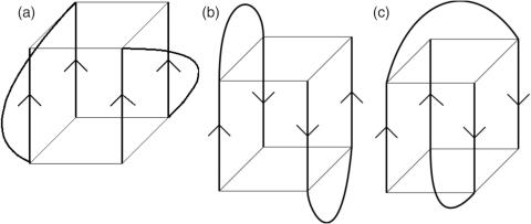
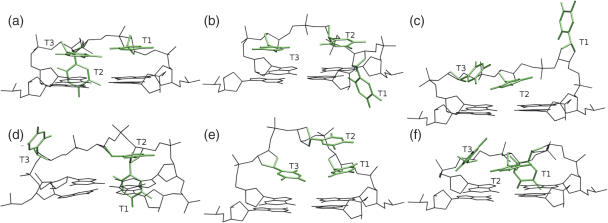
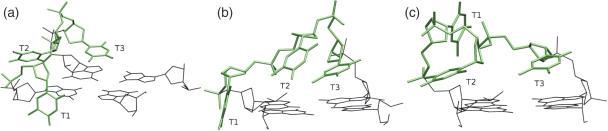
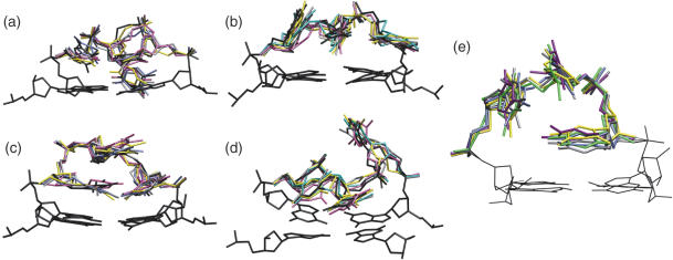
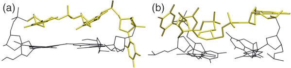
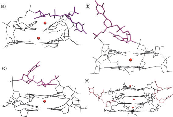
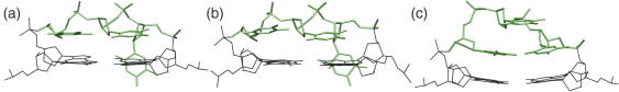
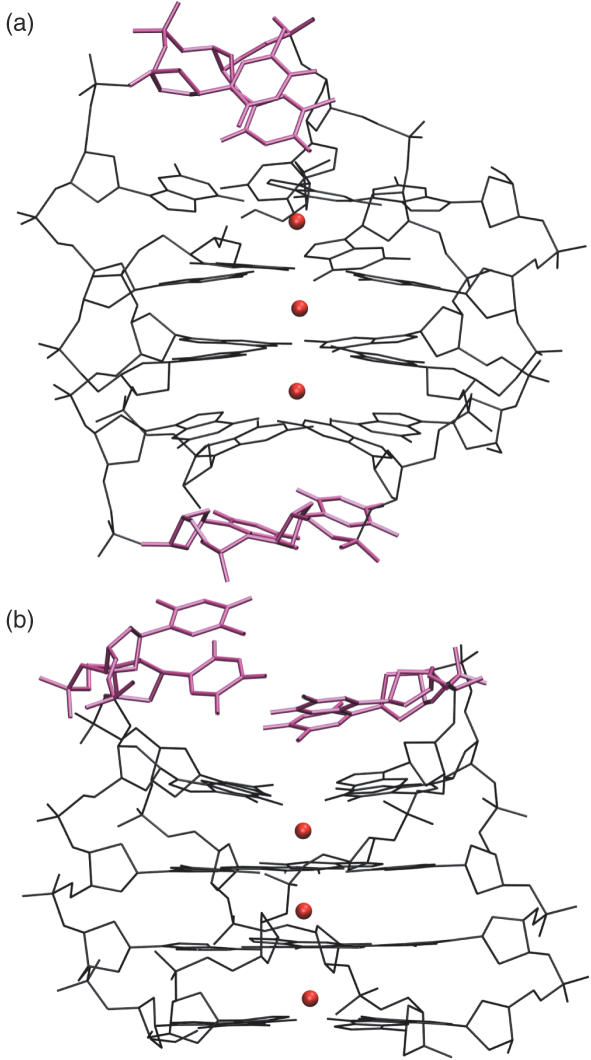
We have used a combination of simulated annealing (SA), molecular dynamics (MD) and locally enhanced sampling (LES) methods in order to predict the favourable topologies and loop conformations of dimeric DNA quadruplexes with T2 or T3 loops. This follows on from our previous MD simulation studies on the influence of loop lengths on the topology of intramolecular quadruplex structures [P. Hazel et al. (2004) J. Am. Chem. Soc., 126, 16 405-16 415], which provided results consistent with biophysical data. The recent crystal structures of d(G4T3G4)2 and d(G4BrUT2G4) (P. Hazel et al. (2006) J. Am. Chem. Soc., in press) and the NMR-determined topology of d(TG4T2G4T)2 [A.T. Phan et al. (2004) J. Mol. Biol., 338, 93-102] have been used in the present study for comparison with simulation results. These together with MM-PBSA free-energy calculations indicate that lateral T3 loops are favoured over diagonal loops, in accordance with the experimental structures; however, distinct loop conformations have been predicted to be favoured compared to those found experimentally. Several lateral and diagonal loop conformations have been found to be similar in energy. The simulations suggest an explanation for the distinct patterns of observed dimer topology for sequences with T3 and T2 loops, which depend on the loop lengths, rather than only on G-quartet stability.


Figures

Figure 1
Figure 1
Schematic diagrams of the (a) parallel, (b) lateral and (c) diagonal loop dimeric G-quadruplexes which were simulated in this work.
Figure 2
Figure 2
Structures with lateral T3 loops over the wide quadruplex groove, generated during SA runs. (a) T3-Lw-1, (b) T3-Lw-2, (c) T3-Lw-3, (d) T3-Lw-4, (e) T3-Lw-5 and (f) T3-Lw-6. The loop bases are shown in green and a G-quartet in black. Lw indicates a lateral loop over the wide quadruplex groove, as opposed to the narrow groove (Ln).
Figure 3
Figure 3
Structures with lateral T3 loops over the narrow quadruplex groove, generated using SA. (a) T3-Ln-1, (b) T3-Ln-2 and (c) T3-Ln-3.
Figure 4
Figure 4
T3 lateral loop final structures after LES simulations. (a) T3-Lw-1 loop 1 and (c) 2 after 4 ns LES simulation, (b) T3-Lw-2 loop 1 and (d) loop 2 after 2.7 ns LES simulation and (e) T3-Ln-1 loop 1 after 4 ns LES simulation. The five LES copies are shown overlapped in each case. A G-quartet is also shown in black.
Figure 5
Figure 5
T3 diagonal loop MD simulation. (a) Stable loop conformation averaged over the final 2 to 4 ns of MD and (b) final structure of the unstable loop, which formed within the last 200 ps of the simulation.
Figure 6
Figure 6
T2 final loop conformations after 4 ns MD simulation (400 ps only for the diagonal loop). (a) Diagonal T2 loop, (b) lateral T2 loop over the narrow groove, (c) lateral T2 loop over the wide groove and (d) parallel T2 loops. The loops are shown in purple, and G-quartets in black, channel K+ ions are shown as red spheres.
Figure 7
Figure 7
MD simulations of (a) d(G4T3G4)2. X-ray structure T3 loop conformation averaged over 2 to 4 ns (b) NMR structure T3 loop conformation averaged over 2 to 4 ns and (c) most favourable predicted T3-Lw-5 loop averaged over 1 to 2 ns MD.
Figure 8
Figure 8
(a) Head-to-tail and (b) head-to-head quadruplex structures with two T2 loops shown in purple. The complete quadruplexes are shown, with K+ channel ions in red. These structures have been averaged over the final 2 ns of the 4 ns simulations.

Similar articles

Cited by

References

    1. Parkinson G.N., Lee M.P.H., Neidle S. Crystal structure of parallel quadruplexes from human telomeric DNA. Nature. 2002;417:876–880. - PubMed
    1. Phan A.T., Patel D.J. Two-repeat human telomeric d(TAGGGTTAGGGT) sequence forms an interconverting parallel and antiparallel G-quadruplexes in solution: distinct topologies, thermodynamic properties, and folding/unfolding kinetics. J. Am. Chem. Soc. 2003;125:15021–15027. - PMC - PubMed
    1. Ambrus A., Chen D., Dai J., Jones R.A., Yang D. Solution structure of the biologically relevant G-quadruplex element in the human c-myc promoter. Implications for G-quadruplex stabilization. Biochemistry. 2005;44:2048–2058. - PubMed
    1. Phan A.T., Modi Y.S., Patel D.J. Propeller-type parallel-stranded G-quadruplexes in the human c-myc promoter. J. Am. Chem. Soc. 2004;126:8710–8716. - PMC - PubMed
    1. Zhan N., Phan A.T., Patel D.J. (3+1) assembly of three human telomeric repeats into an asymmetric dimeric G-quadruplex. J. Am. Chem. Soc. 2005;127:17277–17285. - PMC - PubMed

Publication types